Webs and Lakes in the Heart: The Forgotten Image
© 2025 HMP Global. All Rights Reserved.
Any views and opinions expressed are those of the author(s) and/or participants and do not necessarily reflect the views, policy, or position of the Journal of Invasive Cardiology or HMP Global, their employees, and affiliates.
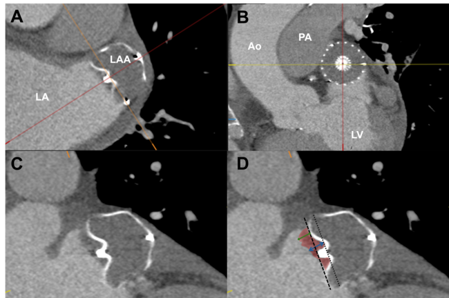
A 50-year-old woman presented to our center with complaints of breathlessness on exertion for the last 2 years. On evaluation, she was diagnosed with rheumatic heart disease with severe calcific mitral stenosis (mitral valve area of 0.9 cm2). The patient was in atrial fibrillation at the time of presentation.
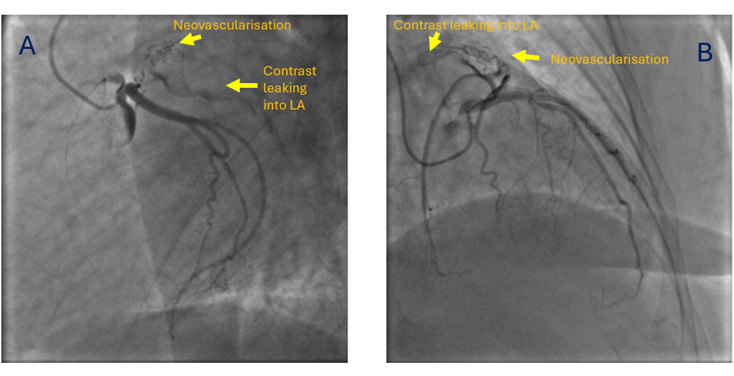
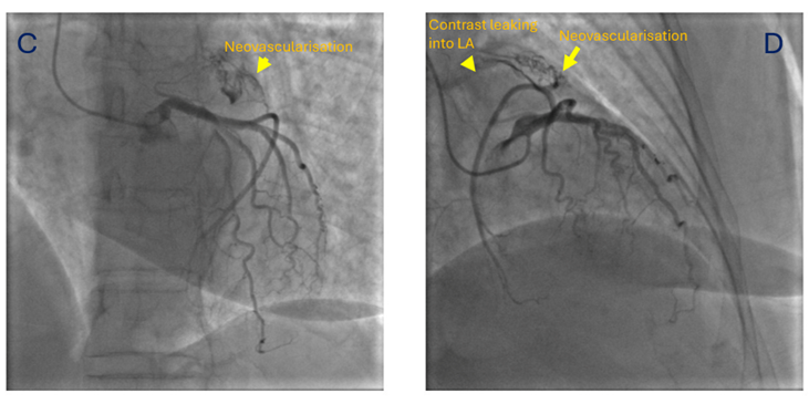
She was taken up for a preoperative coronary angiography with a plan for a subsequent mechanical mitral valve replacement. The coronary angiogram revealed normal epicardial coronary arteries. However, the atrial branch of the left circumflex artery was seen giving small branches to a left atrial appendage (LAA) clot (neovascularization), with contrast leaking into the left atrium (LA) from these neovessels (Figure, Video). A transesophageal echocardiogram later confirmed the LAA clot. The patient was subsequently planned for a mechanical mitral valve replacement and LAA clot removal.
In a previous study from our center, which included 81 patients with rheumatic mitral stenosis, angiography predicted thrombosis by demonstrating neovascularity, manifesting as a bunch of small vessels arising from the circumflex branch of the left coronary artery and terminating in a network of smaller vascular channels, with a blush of contrast medium consolidating into small lakes.1 For recognizing thrombi in the LA or LAA, selective left coronary angiogram demonstrated sensitivity and specificity of 72.7% and 92.7%, respectively, and an 88.8% predictive value.1
Affiliations and Disclosures
Shivam Arora, MD1; Neha Chopra, MD, DM2; Shitij Chaudhary, MD, DM1
From the 1Department of Cardiology, All India Institute of Medical Sciences, New Delhi, India; 2Department of Cardiology, VMMC and Safdarjung Hospital, New Delhi, India.
Disclosures: The authors report no financial relationships or conflicts of interest regarding the content herein.
Consent statement: The authors confirm that informed consent was obtained from the patient for the intervention described in the manuscript and to the publication thereof, including any and all images.
Address for correspondence: Shitij Chaudhary, MD, DM, All India Institute of Medical Sciences, New Delhi 110029, India. Email: shitijchaudhary94@gmail.com
References
1. Sharma S, Kumar MV, Reddy VM, Kaul U, Rajani M, Venugopal P. Left coronary angiography in the pre-operative diagnosis of thrombosis of the left atrium or its appendage in rheumatic mitral stenosis. Clin Radiol. 1990;42(3):188-191. doi:10.1016/s0009-9260(05)81931-2