Clinical Videos
Progression and Patterns of Radial Artery Calcification: Insights From Optical Coherence Tomography
07/15/2025
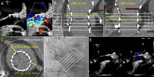
Video 1. Baseline optical coherence tomography cine demonstrated calcium in Figure 1A, but not in Figure 1D.
Video 3. Follow-up optical coherence tomography cine showed calcium progression in Figure 1A′ and 1D′.
Video 4. Follow-up optical coherence tomography cine showed calcium progression in Figure 1B′ and new calcium in Figure 1C′.
© 2025 HMP Global. All Rights Reserved.
Any views and opinions expressed are those of the author(s) and/or participants and do not necessarily reflect the views, policy, or position of the Journal of Invasive Cardiology or HMP Global, their employees, and affiliates.