

Percutaneous Management of a Vascular Stent Graft Malfunction
© 2025 HMP Global. All Rights Reserved.
Any views and opinions expressed are those of the author(s) and/or participants and do not necessarily reflect the views, policy, or position of the Journal of Invasive Cardiology or HMP Global, their employees, and affiliates.
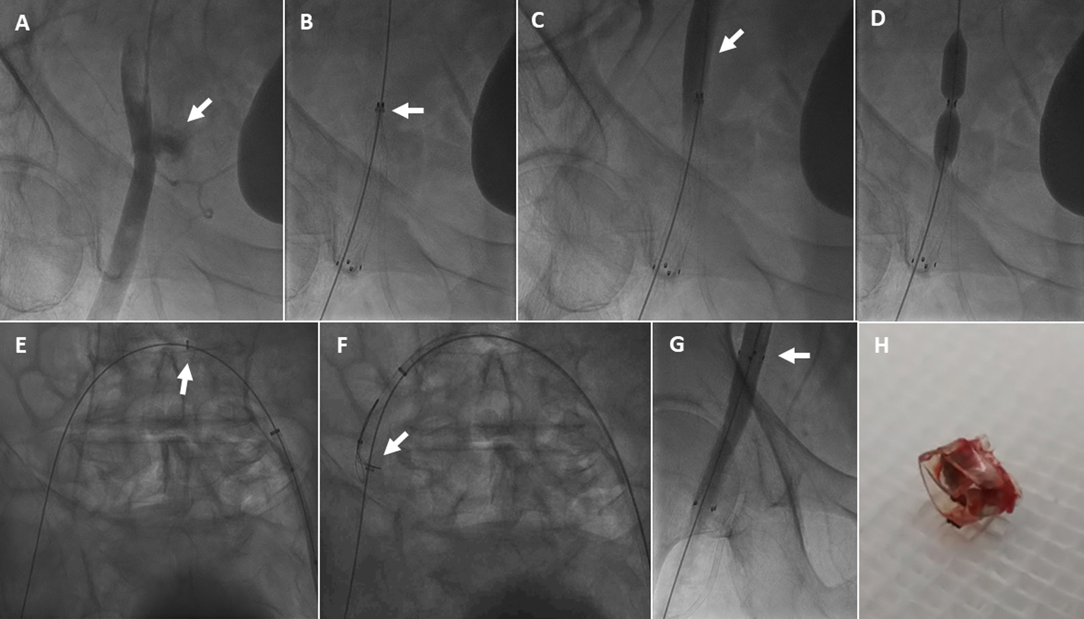
A 69-year-old diabetic man with morbid obesity underwent elective angioplasty for a chronic total occlusion of the left anterior descending coronary artery via right radial (7F) and right femoral (8F) access. Following successful stenting, the right common femoral artery (CFA) was closed using a Perclose ProStyle device (Abbott). The patient became hypotensive, and angiography via radial access revealed bleeding at the right CFA as a result of ProStyle failure (Figure A).
A 10F Flexor Check-Flo sheath (Cook Medical) was advanced via the contralateral left CFA. A 12 × 60-mm Fluency Plus Endovascular Stent Graft (BD) was deployed to seal the perforation in the right CFA. During deployment, a radiopaque ring-like component remained attached to the proximal edge of the stent graft, preventing expansion (Figure B( and leading to arterial occlusion (Figure C). Balloon angioplasty with a 10.0 × 40-mm POWERFLEX Pro balloon (Cordis) failed to expand the stent or displace the ring (Figure D). After balloon deflation and withdrawal, the ring migrated proximally over the guidewire (Figure E, Video 1), allowing proximal expansion of the stent graft. Retrieval attempts using a 4.0 × 40-mm Mustang balloon (Boston Scientific) were unsuccessful. The ring was ultimately captured and retrieved using a 12 x 20-mm EN Snare Endovascular System (Merit Medical) (Figure F, Video 2). Final angiography confirmed complete stent-graft expansion and restored blood flow (Figure G, Video 3). The left CFA was successfully closed with a Perclose ProStyle device.
We describe a previously undescribed complication: incomplete deployment of a stent graft, leading to vascular occlusion. Although the source of the detached component could not be confirmed (Figure H), its radiopacity and morphology suggest it was part of the delivery system. Prompt percutaneous retrieval using balloon and snare techniques resolved this complication without need for vascular surgery.
Affiliations and Disclosures
Ibrahim Naoum, MD1; Hashem Hayeq, MD2; Amnon Eitan, MD1; Moataz Tarabih, MD1; Ronen Jaffe, MD1
From the Departments of 1Cardiology and 2Vascular and Endovascular Surgery, Lady Davis Carmel Medical Center, and the Ruth and Bruce Rappaport Faculty of Medicine, Technion–Israel Institute of Technology, Haifa, Israel.
Disclosures: The authors report no financial relationships or conflicts of interest regarding the content herein.
Consent statement: The authors confirm that informed consent was obtained from the patient for the intervention described in the manuscript and for the publication thereof, including any and all images.
Address for correspondence: Ronen Jaffe, MD, Department of Cardiovascular Medicine, Lady Davis Carmel Medical Center, 7 Michal St, Haifa 34362, Israel. Email: jafferonen@gmail.com


