

Acute Allergic Blepharoconjunctivitis During Percutaneous Coronary Intervention
© 2025 HMP Global. All Rights Reserved.
Any views and opinions expressed are those of the author(s) and/or participants and do not necessarily reflect the views, policy, or position of the Journal of Invasive Cardiology or HMP Global, their employees, and affiliates.
A 44-year-old man presented with typical angina on minimal exertion (New York Heart Association Class III) for the last 2 weeks, which had gradually worsened to rest angina. On examination, there were no significant findings; the 12-lead electrocardiography and 2-dimensional transthoracic echocardiography were within normal limits. The patient was taken for coronary angiogram, which revealed significant stenosis of 90% in the mid-left anterior descending artery (LAD) with nonobstructive mild plaquing in the remaining coronary arteries.
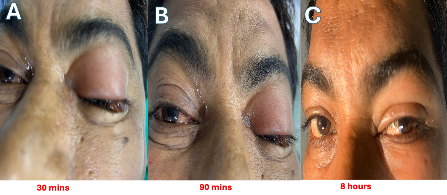
During percutaneous coronary intervention to the LAD, the patient complained of itching and discomfort in both eyes, with more significant symptoms in his left eye. On inspection, there was redness and swelling in both eyes with more remarkable changes in the left eye compared with the right. The swelling in the eyelids progressed significantly, almost completely occluding the left eye. We suspected an acute allergic contrast reaction and administered intravenous antihistamines and hydrocortisone. The swelling started within 30 minutes of administration of contrast (iohexol), peaked at 90 minutes following the first angiography shot, and stopped progressing once intravenous drugs were administered (Figure).
As the patient responded very well to the treatment, a diagnosis of isolated allergic blepharoconjunctivitis secondary to contrast was made. The swelling subsided slowly over 8 hours with complete normalization of the congestion and swelling by next morning.
Affiliations and Disclosures
Devesh Kumar, MD, MRCP, DM1; Sourabh Agstam, MD, DM, MRCP2
From the 1Department of Cardiology, Vardhman Mahavir Medical College and Safdarjung Hospital, New Delhi, India; 2Department of Cardiology, All India Institute of Medical Sciences, New Delhi, India.
Disclosures: The authors report no financial relationships or conflicts of interest regarding the content herein.
Consent statement: The authors confirm that informed consent was obtained from the patient for the clinical photographs described in the manuscript and for the publication of thereof.
Address for correspondence: Sourabh Agstam, MD, MRCP, DM, Department of Cardiology, 7th floor, Cardio-Thoracic Centre, All India Institute of Medical Sciences, New Delhi 110029, India. Email: sourabhagstam@gmail.com; X: @AgstamSourabh


