Finding the Optimal Pacing Threshold and Avoiding Multiple Deployments of the Micra Device Using Temporary Venous Pacing Mapping
Video Supplement to "Finding the Optimal Pacing Threshold and Avoiding Multiple Deployments of the Micra Device Using Temporary Venous Pacing Mapping" (Clinical Image).
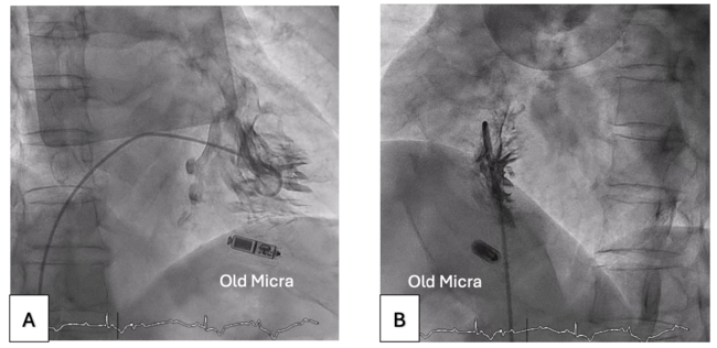
Video 1B. Right ventriculogram via pigtail catheter in the left anterior oblique view. Consistent fluoroscopic angles and table height were maintained after the right ventriculogram.
Video 2A. The optimal pacing site was identified by temporary venous pacing in the right anterior oblique view.
Video 2B. The optimal pacing site was identified by temporary venous pacing in the left anterior oblique view. These images served as the fluoroscopic roadmap for deployment of the new Micra AV (Medtronic).
Video 3A. The new Micra AV (Medtronic) was targeted at the temporary venous pacing-identified site in the right anterior oblique view.
© 2025 HMP Global. All Rights Reserved.
Any views and opinions expressed are those of the author(s) and/or participants and do not necessarily reflect the views, policy, or position of the Journal of Invasive Cardiology or HMP Global, their employees, and affiliates.