

Thrombotic Complication After a Double-Barrel Configuration Percutaneous Coronary Intervention in a Chronic Total Occlusion
© 2025 HMP Global. All Rights Reserved.
Any views and opinions expressed are those of the author(s) and/or participants and do not necessarily reflect the views, policy, or position of the Journal of Invasive Cardiology or HMP Global, their employees, and affiliates.
A 63-year-old man with stable angina was scheduled for elective percutaneous coronary intervention of a chronic total coronary occlusion of the right coronary artery (RCA) with epicardial bridging collaterals from the left circumflex artery to the posterior descending artery. He had previously received stents in the mid-RCA.
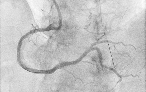
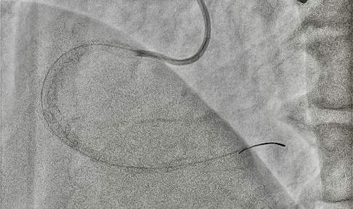
A Convey CLS 4 6F guiding catheter (Boston Scientific) was engaged in the left main coronary artery, and an Amplatz left 1 7F was engaged in the ostium of the RCA. Starting from the antegrade with a Gladius EX guidewire (Asahi) and a Turnpike Spiral microcatheter (Teleflex), a dissection reentry was attempted. Τhe Gladius EX, the Gaia Next 2 (Asahi), the Confianza Pro 12 (Asahi), and the Hornet 14 (Boston Scientific) were used consecutively, passing inside the outer layer of the vessel (adventitia), next to the old stent, and reentering in the distal part of the RCA. In order to reenter the true lumen before the bifurcation, a Stingray balloon (Boston Scientific) was used. A Gaia Next 3 was advanced inside the Stingray, successfully reentering the lumen. Afterwards, balloon inflation was performed throughout the vessel and, finally, 4 stents were deployed with a good angiographic result and Thrombolysis in Myocardial Infarction-III flow (Figures 1 and 2, Videos 1 and 2). The patient was discharged the next day.
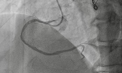
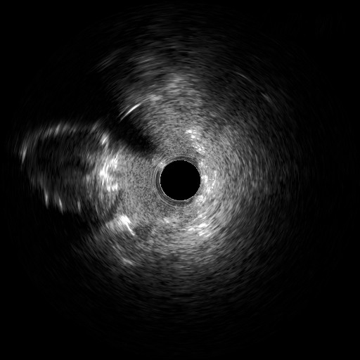
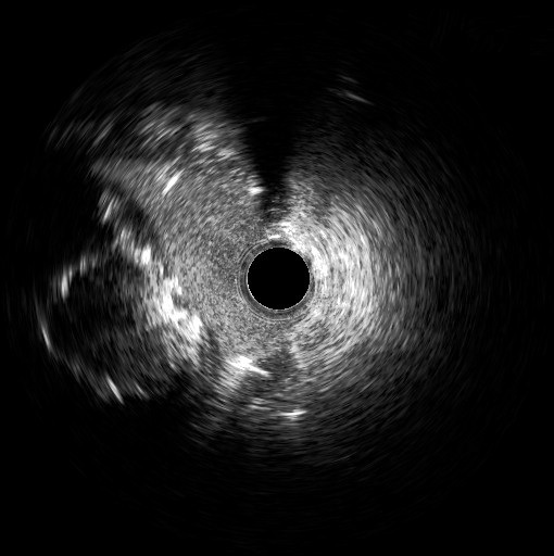
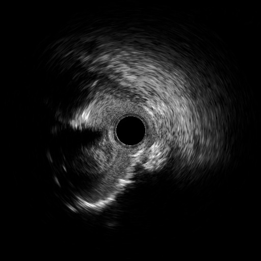
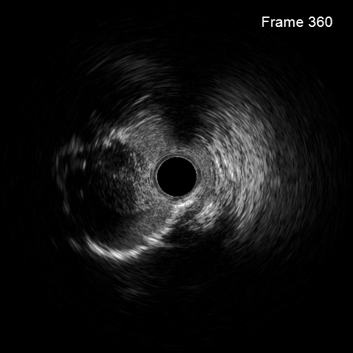
Four days later, the patient was admitted to the cardiology department because of exertional dyspnea. Because of the recent revascularization of the RCA, a new coronary angiography was performed. The angiography of the left system revealed no changes, but the RCA unveiled a nimble formation in the proximal part of the stented vessel (Figure 3, Video 3). An intravascular ultrasound was performed, which displayed the double-barrel configuration (Figures 4 and 5, Video 4) and the presence of a thrombus (Figures 6 and 7, Video 5). It was directly stented with an excellent angiographic result (Video 6). Two days postoperative, the patient was asymptomatic and discharged.
Affiliations and Disclosures
Panagiotis Varelas, MD, MSc1; Konstantinos Manousopoulos, MD, PhD1; Konstantinos Filippou, MD, MSc1; Dimitrios Karelas, MD, MSc1; Ioannis Papadopoulos, MD1; Alexandre Avran, MD2; Ioannis Tsiafoutis, MD, PhD1
From the 1Red Cross Hospital, Korgialeneio-Benakeio, Athens, Greece; 2Valenciennes Hospital, Valenciennes, France.
Disclosures: The authors report no financial relationships or conflicts of interest regarding the content herein.
Consent statement: The authors confirm that informed consent was obtained from the patient for the intervention described in the manuscript and to the publication of this manuscript, including any and all images.
Address for correspondence: Panagiotis Varelas, MD, MSc, Erithrou Stavrou 1 & Athanasaki Ioanni, Ampelokipoi, Athens 11526, Greece. Email: pvarelas@outlook.com


