

Progression and Patterns of Radial Artery Calcification: Insights From Optical Coherence Tomography
© 2025 HMP Global. All Rights Reserved.
Any views and opinions expressed are those of the author(s) and/or participants and do not necessarily reflect the views, policy, or position of the Journal of Invasive Cardiology or HMP Global, their employees, and affiliates.
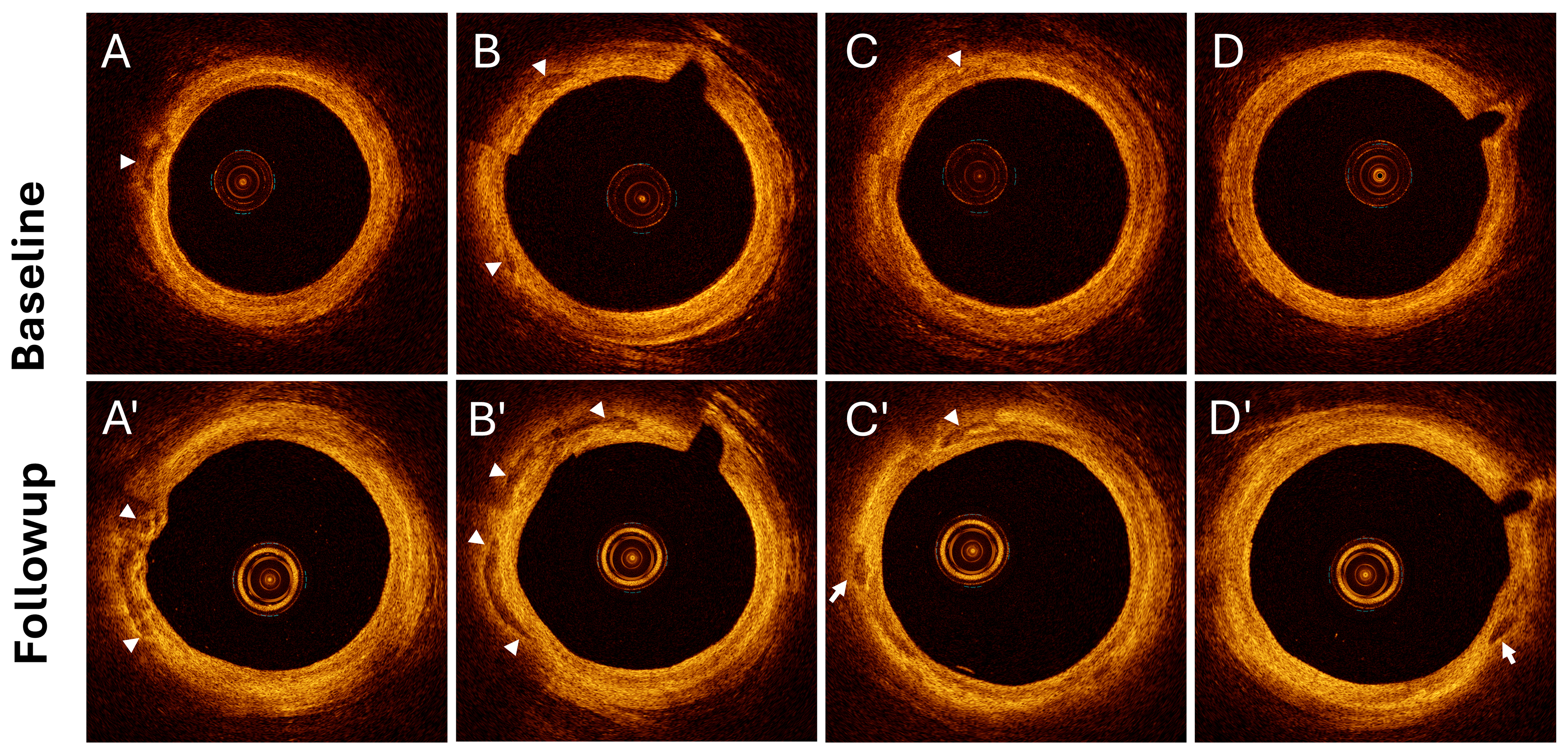
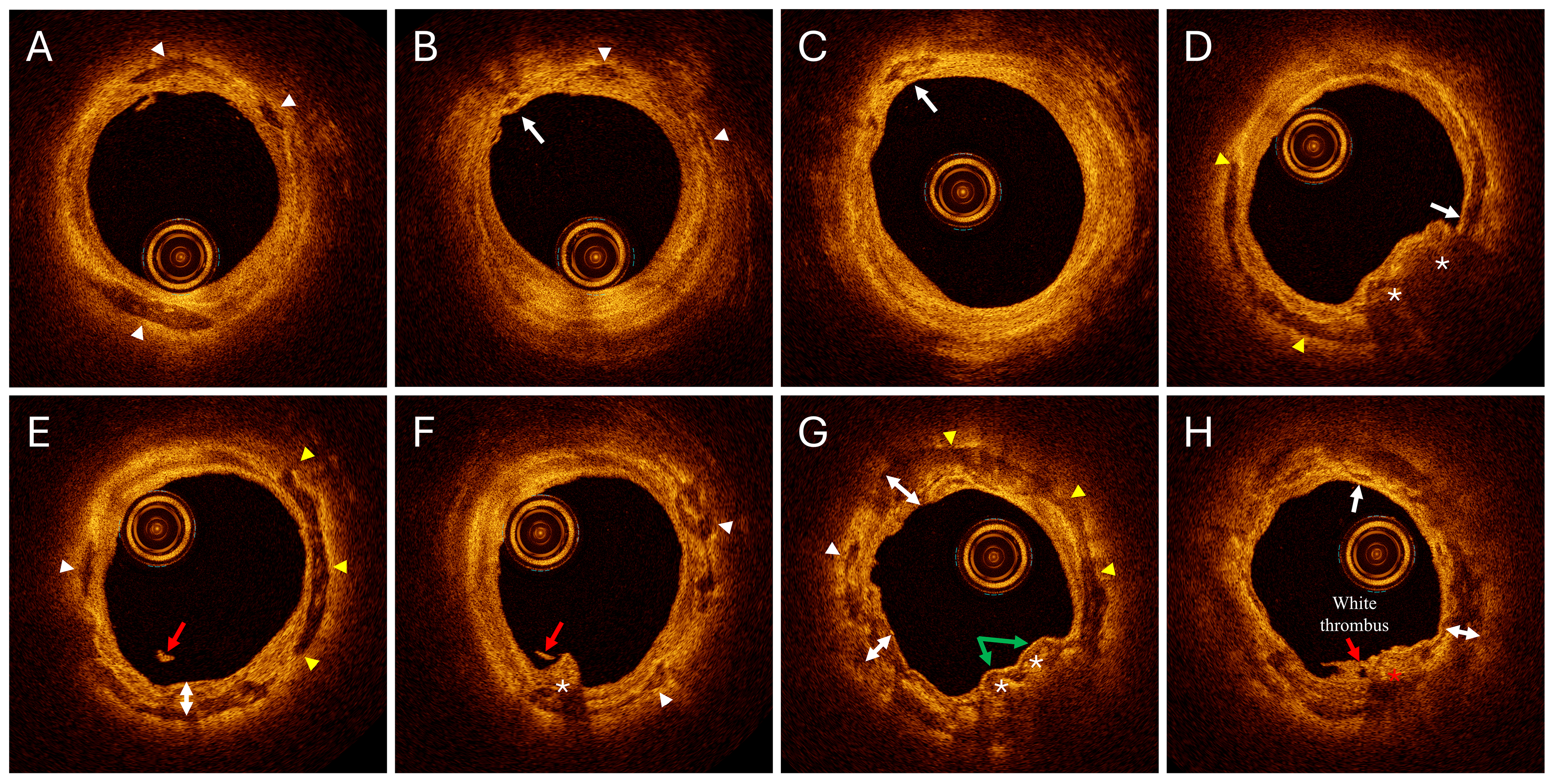
A 72-year-old man with a history of hypertension and diabetes mellitus underwent coronary intervention via distal radial artery (RA) access, guided by optical coherence tomography (OCT), for recurrent angina. A follow-up 18-cm OCT pullback of the RA was performed immediately post-procedure and compared with baseline 15-cm OCT images, which had been obtained 5 years earlier via radial access. The 2025 OCT revealed marked progression of RA calcification (Figure 1, Videos 1-4). Furthermore, in distal RA regions not included in the baseline scan, OCT revealed diverse calcification phenotypes (Figure 2, Video 5).
This case uniquely illustrates the dynamic nature of RA calcification progression and identifies calcification patterns analogous to those commonly observed in coronary arteries.
Affiliations and Disclosures
Saiying He, MD; Jia Zhou, MD; Hao Liu, MD; Jincheng Guo, MD
From the Department of Cardiology, Beijing Luhe Hospital, Capital Medical University.
Disclosures: The authors report no financial relationships or conflicts of interest regarding the content herein.
Consent statement: The authors confirm that informed consent was obtained from the patient for the study and intervention described in the manuscript and to the publication of their data including any and all images.
Address for correspondence: Jincheng Guo, MD, Department of Cardiology, Beijing Luhe Hospital, Capital Medical University, Tongzhou District, Beijing 101149, China. Email: guojcmd@126.com


