

Congenital Coronary Artery-Right Ventricular Multiple Microfistulas
© 2025 HMP Global. All Rights Reserved.
Any views and opinions expressed are those of the author(s) and/or participants and do not necessarily reflect the views, policy, or position of the Journal of Invasive Cardiology or HMP Global, their employees, and affiliates.
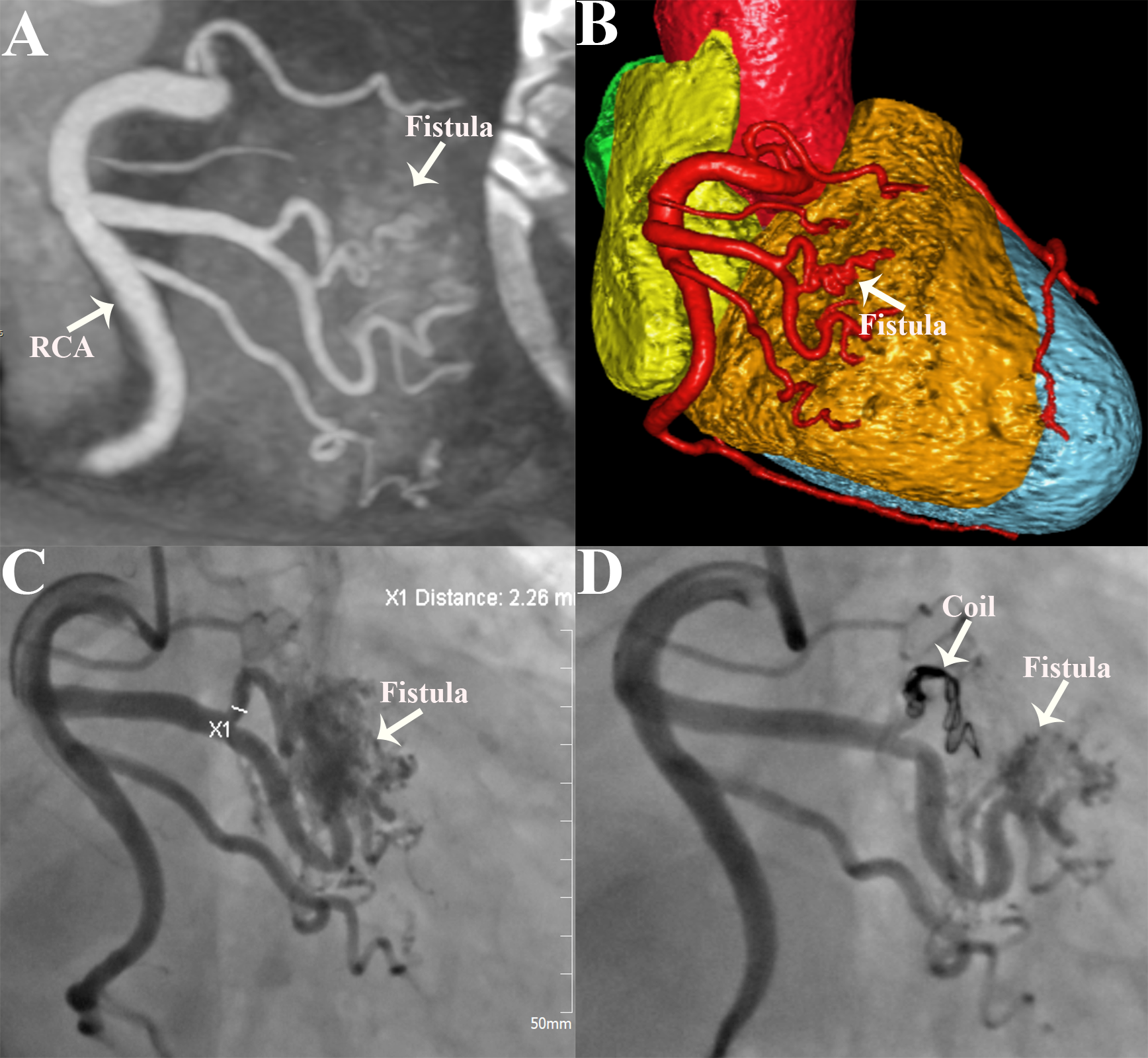
A 65-year-old man presented with intermittent chest tightness and shortness of breath for 8 months. The physical examination and laboratory tests were unremarkable. The electrocardiogram revealed sinus rhythm and inverted T-waves in leads II, III, and aVF. Transthoracic echocardiography showed an enlarged left heart and mild left ventricular hypodiastolic dysfunction. Coronary computed tomography angiography (CCTA) showed multiple microfistula between the multiple branches of the right coronary artery (RCA) and right ventricle (Figures A and B). Coronary angiography (CAG) demonstrated coronary artery-right ventricular multiple microfistulas (CA-RVMMFs) arising from the branches of the RCA (Figure C, Video 1).
The patient was treated by embolizing a large fistula vessel with an interlock coil (Fibered IDC; Boston Scientific) measuring 5 mm x 15 cm. Postoperative color Doppler flow imaging and CAG images revealed that the coronary fistula had been successfully embolized and the amount of shunting was significantly decreased (Figure D, Video 2). The postoperative course was uneventful, and the patient was discharged after 3 days. At the 6-month follow‑up, the patient's symptoms were significantly relieved.
Congenital CA-RVMMFs are rare anomalies, and the clinical and hemodynamic sequelae are incompletely understood.1 They result from the failure of obliteration of intertrabecular embryonic sinusoids and may cause angina pain or myocardial ischemia through the coronary steal mechanism.2 The characteristics of CA-RVMMFs during selective CAG are the visualization of small vessels interposed between the epicardial coronary vessels and the heart cavity.3 The optimal management of CA-RVMMFs is controversial, with no established guidelines. Options include percutaneous intervention, particularly in symptomatic patients or those with existing myocardial ischemia. Our case highlights the crucial role of CCTA and CAG in the accurate diagnosis and guidance of clinical treatment.
Affiliations and Disclosures
Leizhi Ku, MD1; Yuhang Wang, MD1; Zheng Liu, MD1; Xiaojing Ma, PhD2
From the Departments of 1Radiology and 2Echocardiography, Wuhan Asia Heart Hospital, Wuhan University of Science and Technology, Hankou District, Wuhan, P.R. China.
Dr Ku and Dr Wang contributed equally to the article.
Disclosures: The authors report no financial relationships or conflicts of interest regarding the content herein.
Funding: This work was funded by the Wuhan Clinical Medical Research Center for Cardiovascular Imaging (CMRC202307).
Consent statement: Informed consent was obtained from the patient for this study.
Statement of artificial intelligence (AI): All authors declare not to have used artificial AI or AI-assisted technologies in the production of submitted work.
Address for correspondence: Xiaojing Ma, PhD, Department of Echocardiography, Wuhan Asia Heart Hospital Affiliated Wuhan University of Science and Technology, No. 753 Jinghan Road, Hankou District, Wuhan 430022, P.R. China. Email: klz1534292102@163.com; X: @klz13657254286
References
1. Stierle U, Giannitsis E, Sheikhzadeh A, Potratz J. Myocardial ischemia in generalized coronary artery-left ventricular microfistulae. Int J Cardiol. 1998;63(1):47-52. doi:10.1016/s0167-5273(97)00280-5
2. Abubakar H, Ahmed AS, Adam O, Yassin AS. Three vessel coronary artery-left ventricular multiple micro-fistulas: a rare angiographic finding. Oxf Med Case Reports. 2018;2018(8):omy053. doi:10.1093/omcr/omy053
3. Doğan M, Sunman H, Akyel A, et al. Koroner arter ile sol ventrikül arasındaki mikrofistüllerin sıklığı ve klinik özellikleri: tek merkez deneyimi [Prevalence and clinical features of microfistulas between the coronary artery and left ventricle: single-center experience]. Turk Kardiyol Dern Ars. 2014;42(4):332-338. doi:10.5543/tkda.2014.25936


