Case Report
Unintended Iatrogenic Creation of an Internal Thoracic Artery to Anterior Coronary Vein Bypass Graft with Subsequent Reoperation
September 2004
Coronary artery bypass surgery is now a common operation in major tertiary centers with over 300,000 surgeries performed annually in the United States. Despite the refinement of the procedure and high volume experience of most surgeons, mistakes are still possible. Many patients have significant comorbid conditions and on the average the surgery carries an operative mortality risk of approximately 3%. There are many potential complications of the operation including the inability to properly identify the appropriate coronary vessel. Inadvertent anastamosis of a bypass conduit to a coronary vein was first reported in 1975.1 We describe the first reported case of reoperation to correct an inappropriately placed LITA to a coronary vein with preservation of the LITA conduit and reimplantation into a coronary artery.
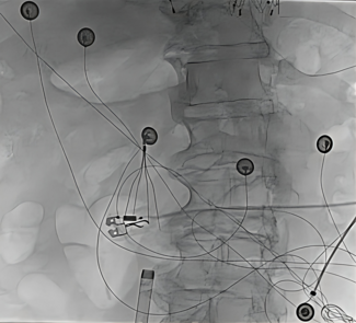
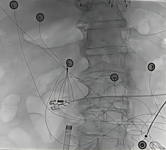
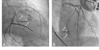
Case Report. The patient is a 73-year-old man who had a myocardial infarction approximately 17 years ago. He was initially treated medically, but several months later he became unstable and required four-vessel coronary bypass surgery. The intention was to place the LITA to the LAD coronary artery and to place three dedicated reversed autologous saphenous vein grafts to the right coronary artery (RCA), marginal branch, and diagonal branch of the LAD. The patient recovered from surgery and was clinically stable for many years. He enjoyed an active lifestyle and was without cardiac symptoms. Several years before this presentation he had an echocardiogram that showed severe left ventricular (LV) systolic dysfunction. He was treated with appropriate medications and remained asymptomatic. He subsequently developed exertional chest discomfort radiating to both arms. The cardiac exam revealed no murmur. He underwent elective catheterization where he was found to have severe native vessel coronary artery disease and severe LV dysfunction with the ejection fraction (EF) estimated at 20%. Two of the vein grafts were 100% occluded proximally, and the vein graft to the RCA was patent. Contrast injection in the LITA graft showed no lesions and good flow. After the insertion of the graft the flow continued superiorly back to the base of the heart and then to the coronary sinus (Figures 1 and 2). There was no flow toward the anteroapex of the heart. Because of these findings at catheterization, repeat coronary artery bypass grafting (CABG) was recommended.
The operative risk was felt to be high because of repeat mediansternotomy and severe left ventricular dysfunction. An additional concern was the lack of ideal conduit since the LITA had already been used as well as prior vein grafts. The patient had repeat CABG which was uncomplicated. The distal insertion of the LITA to the anterior coronary vein was interrupted and the LITA was repositioned and anastamosed to the ramus intermedius. A free radial graft was used to bypass the LAD, and reversed autologous saphenous vein grafts were used to bypass the posterior descending artery and the diagonal branch of the LAD.
After recovery, the patient returned to his vigorous lifestyle. Follow up echocardiography at 6 months post-surgery showed improved LV function with the EF estimated at 30–35%.
Discussion. In the early 1970s, operations were done for the treatment of myocardial ischemia to intentionally shunt flow from the aorta to the coronary veins.2,3 These operations fell out of favor as the standard continued to be the use of the coronary artery as the distal target. The rare complication of an iatrogenic shunt by the inadvertent insertion of the conduit to a coronary vein was first reported in 1975.1 In that case, the diagnosis was suspected because of the finding of a postoperative continuous murmur. Since then, there have been dozens of cases in the literature. Presumably there are many cases that were never reported or never diagnosed. There are no reports of direct mortality from this misadventure, but it is possible that this complication was present in some cases of unexpected perioperative death. While the possibility of mortality is unknown, certainly there is morbidity. It is difficult to accurately define the morbidity associated with such a rare complication, but theoretic possibilities include incomplete revascularization with ongoing ischemia, the creation of a left to right shunt, waste of the arterial conduit, and the possible need for subsequent reoperation. Endocarditis has not been reported, but has been raised as a possible concern. Treatment options have included conservative observation, percutaneous occlusion, and open surgical repair.4
It is interesting to consider why bypassing a coronary vein rather than the artery might be possible. Typically, the coronary artery is a thick walled vessel that often can be distinguished from the vein by palpation because of plaque. Often the artery is quite thin with no plaque distally making differentiation between artery and vein difficult. The artery may be identified by observing its location and by finding the branch vessels and tracing them back to the main epicardial coronary arteries. Red, oxygenated, blood and pulsatile flow can be used to identify the artery before application of the cross clamp. During bypass operation the surgeon may have incomplete exposure or not take time to carefully identify the correct vessel. Redo surgery often obscures the topical coronary anatomy. Arteries that take an intramyocardial course or are thin walled may be more difficult to correctly identify. Generally, veins are thin walled, contain dark deoxygenated blood, and are on the surface of the heart.
Our patient's operative risk was high due to his severe LV dysfunction. The LITA was used at the first surgery, but we felt that it should be salvaged, in order to utilize an arterial conduit. There was concern about damage to the LITA at repeat sternotomy. The LITA could have been ligated with a new vein or arterial graft created to the LAD. Another option was to interrupt its insertion and reattach the distal vessel to the LAD. The LITA could also be mobilized and anastamosed to the ramus intermedius (which we did), or harvested as a free LITA conduit. There was consideration given to use of the right internal thoracic artery (RITA), but it was not used because of adequate conduit elsewhere. Because of the length of the LITA, and the concern that it would not reach the LAD, the radial artery was harvested as a free conduit to bypass the LAD. The distal insertion of the LITA was taken down and the LITA anastamosed to the ramus intermedius artery.
Conclusion. Unintended iatrogenic internal thoracic artery to coronary vein shunt is a rare complication of coronary artery bypass surgery. The mortality seems to be low, but there is associated morbidity. Surgical vigilance should prevent this complication, however, the diagnosis should be considered if an unexpected continuous murmur is heard postoperatively. There is a spectrum of reasonable choices in the management of this condition.
1. Vieweg W, Folkerth T, Hagan A. Saphenous vein graft from aorta to coronary vein with production of continuous murmur. A complication of coronary artery bypass surgery. Chest 1975;68:377–379.
2. Sallam I and Kolff W. A new surgical approach to myocardial revascularization — Internal mammary artery to coronary vein anastomosis. Thorax 1973;28:613–616.
3. Lichtlen P. The aorto-to-coronary vein bypass graft: Post-operative clinical and functional evaluation. J Cardiovasc Surg 1974;15:163–173.
4. Calkins J, Talley J, Kim N. Iatrogenic aorto-coronary venous fistula as a complication of coronary artery bypass surgery: Patient report and review of the literature. Cathet Cardiovasc Diagn 1996;37:55–59.


