Case Report
uccessful Bailout Percutaneous Coronary Intervention for Immediate Surgical Complication
September 2004
Coronary artery compression can occur during mitral valve replacement with a mechanical prosthesis due to the proximity of the dominant artery to the posterior mitral annulus and the propensity for distortion during anchoring the prosthesis sewing ring. This can result in irreversible myocardial ischemia, infarction and even death post-operatively if there is complete vessel occlusion.1–6 Very few articles have mentioned the treatment of such an injury. We previously reported the first successful treatment of coronary artery compression after mitral valve replacement in a child using a coronary stent.7 We now report the first adult case of complete dominant left circumflex artery occlusion discovered immediately post mitral valve mechanical prosthesis and treated using three drug-eluting stents with excellent results. We propose to consider this less invasive percutaneous bailout approach as an alternative (or hybrid) treatment for such surgical complications in order to avoid exposing the patient to the risk of redo surgery.
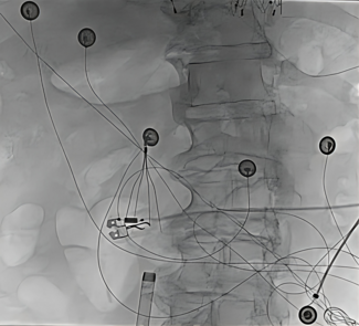
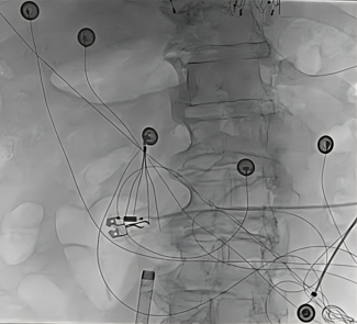
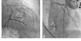
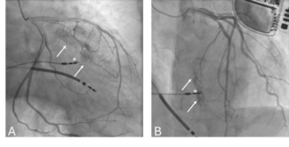
Case report. Our patient was a 36-year-old man with history of rheumatic heart disease. He underwent aortic valve replacement with mechanical St. Jude prosthesis 20 years prior. Recently, he became symptomatic with severe rheumatic mitral regurgitation and underwent mitral valve replacement with a 29 mm CarboMedicics mechanical prosthesis (Sulzer medica prosthetic heart valve, Carbomedics Inc., Austin, Texas). Immediately post-operatively, his 12-lead electrocardiogram showed significant acute ST elevation (acute injury pattern) involving the inferior and posterior leads. Echocardiogram showed severe hypokinesis of the inferior and posterior walls. Thus, the patient was taken emergently to the cardiac catheterization laboratory, where coronary angiography was performed. This revealed normal coronary origins, a left dominant coronary circulation, total occlusion of the middle circumflex artery around the mitral mechanical prosthesis with no flow to the distal area, and normal vessels elsewhere (Figures 1 and 2).
After group discussion of the management options, risks and benefits, we opted to proceed with percutaneous transcatheter intervention to treat this complication. Using a 7 French Left Judkins guiding catheter, 0.014" ACS Hi-Torque Cross-It and 0.014´´ ACS Hi-Torque floppy II wires with MICROGLIDE coating, we successfully crossed the totally occluded segment of the dominant left circumflex coronary artery after several attempts, then passed a 2.5 x 20 mm Tacker balloon (Cordis Corporation, Miami, Florida) dilated up to 12 atmospheres. We established distal flow, but still had phasic compression of the artery around the mitral valve prosthetic ring (Figures 3 and 4). We decided to overcome this mechanical compression using metal scaffolding with coronary stents. We successfully deployed 3 sirolimus-eluting Cypher™ stents (Cordis Corporation). First, we deployed a 2.5 x 18 mm Cypher stent distally at 12 atm, followed by a 2.5 x 18 mm Cypher stent in the mid segment deployed at 12 atm and a 3.0 x 8 mm Cypher stent in the proximal segment deployed at 12 atm. All were deployed with excellent results, with no residual lesions or compression and TIMI 3 flow with normal myocardial blush (Figures 5 and 6). Post-procedure, the patient had complete resolution of his ST elevation and mild elevation of troponin T and CK-MB levels. He was extubated on the following day, remained hemodynamically stable, and was maintained on anticoagulation, antiplatelet therapy, beta-blocker and ACE inhibitor. Repeat echocardiogram a few days later showed near normal wall motion and normal function of both mitral and aortic mechanical prostheses. The patient was seen regularly in the clinic; repeat coronary angiography was done 1 year later and revealed a widely patent left circumflex artery (Figure 7).
Discussion. Coronary artery injury during valve surgery may occur with occlusion of the artery by the prosthetic ring, or by laceration, dissection or suture of the artery.1 The close proximity of the dominant left circumflex artery to the mitral annulus (average, 3–7.5 mm)2 contributes to the risk of iatrogenic injury, especially to the proximal mid segment of the artery during anchoring the prosthetic ring in place. Early recognition and management is crucial to maintain myocardial viability and function. Immediate saphenous vein bypass grafting to the distal left circumflex was the common treatment for such complication (especially for those who could not be weaned from bypass machine).2 We now report a successful new treatment approach via percutaneous catheter intervention and using drug-eluting stents for consideration as an alternative (or hybrid) technique to treat such surgical complications.
1. Roberts WC, Morrow AG. Compression of anomalous left circumflex coronary arteries by prosthetic valve fixation rings. J Thorac Cardiovasc Surg 1969;57:834–838.
2. Virmani R, Chun PK, Parker J, McAllister HA Jr. Suture obliteration of the circumflex coronary artery in three patients undergoing mitral valve operation. Role of left dominant or codominant coronary artery. J Thorac Cardiovasc Surg 1982;84:773–778.
3. Veinot JP, Acharya VC, Bedard P. Compression of anomalous circumflex coronary artery by a prosthetic valve ring. Ann Thorac Surg 1998;66:2093–2094.
4. Cornu E, Lacroix PH, Christides C, Laskar M. Coronary artery damage during mitral valve replacement. J Cardiovasc Surg (Torino) 1995;36:261–264.
5. Morin D, Fischer AP, Sohl BE, Sadeghi H. Iatrogenic myocardial infarction. A possible complication of mitral valve surgery related to anatomical variation of the circumflex coronary artery. Thorac Cardiovasc Surg 1982;30:176–179.
6. Mulpur AK, Kotidis KN, Nair UR. Partial circumflex artery injury during mitral valve replacement: Late presentation. J Cardiovasc Surg (Torino) 2000;41:333–334.
7. Assaqqat MA, Hassan W, Siblini G. Coronary arterial compression treated by stenting after replacement of the mitral valve in a child. J Cardiol Young 2003;13:475–478.


