Case Report
Treatment of Iatrogenic Aortic Dissection by Percutaneous Stent Placement
March 2003
Aortic dissection is a rare but recognized complication of coronary angiography. In early series, angioplasty-induced dissection and other acute events associated with abrupt vessel closure complicated percutaneous transluminal coronary angioplasty in 4–5% of cases.1 The reported frequency of angiography or angioplasty-induced dissection in peripheral vessels varies in the literature. In the series by Gardiner et al., the frequency of dissection due to iliac angioplasty was stated to be 1.3% (3 of 224).2 In the largest published series of 15,500 angiographic procedures conducted in three centers over 6 years, only 6 cases of iatrogenic aortic dissection were identified.3 There was 1 case of anterograde dissection, two cases of retrograde dissection and 3 cases in which the dissection extended in both anterograde and retrograde directions from the injury site on computed tomography (CT) scanning. In contrast to retrograde dissections (extending in opposite direction to blood flow in the true lumen), which reduced or resolved in 1–3 months, anterograde dissections (extending in the direction of blood flow in the true lumen) persisted on CT at 15–27 months.
The duration of patency of an aortic dissection depends on the direction of the dissection. Retrograde dissections frequently decrease or disappear relatively quickly, whereas anterograde dissections often remain patent for a long time. This difference is understandable because blood pressure is pulsatile in an anterograde dissection but not in a retrograde dissection.4 Furthermore, in patients in whom dissection is anterograde with both entry and exit sites, stenting of the distal exit site may worsen the dissection, presumably because the stent at the distal end of the dissection may create high resistance in the dissection channel. Such increased resistance may result in expansion of the dissection channel and closure of the true lumen proximally.5
Intraluminal stents have been shown to be effective in the treatment of iatrogenic iliac dissection.5 The aim of stenting is to eliminate or reduce the dissection flap and to restore luminal patency to a size commensurate with sizes of the vessel segments above and below the stent(s).
We present the case of a woman who suffered an aortic dissection during elective cardiac catheterization via the right femoral artery. This is the first report of aortic dissection, a rare complication of angiography, managed by percutaneous stenting of the entry point.
Case Report. A 57-year-old woman with suspected coronary disease had cardiac catheterization via the femoral approach. A standard right femoral artery puncture was performed and the guidewire passed easily to the mid-aorta. However, there was difficulty advancing the guidewire and the catheter beyond the aortic arch and the procedure was abandoned. Half an hour later, the patient complained of severe back pain and became temporarily hypotensive. She was transferred to our unit, where on arrival, she was pain free but tachycardic (120 beats/minute) and blood pressure was 90/50 mmHg in both arms.
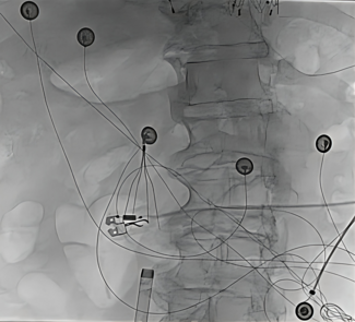
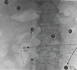
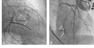
A transesophageal echo revealed a dissection flap commencing in the arch of the aorta. A contrast-enhanced spiral CT scan confirmed aortic dissection, with the entry point in the femoral artery extending to the aortic arch (Figure 1). Initially, the patient was managed conservatively, but she had recurrent transient episodes of severe back pain associated with transient hypotension (75/40 mmHg). A repeat CT scan 8 hours after the procedure showed minor proximal extension of the dissection in the aortic arch and entry site in the right external iliac artery. It was hypothesized that the entry site in the pelvic vessels was responsible for continued filling of the dissection in the thoracic and abdominal component. After discussion, it was decided to close this pelvic vessel entry flap by placement of a self-expanding metallic stent. Under fluoroscopic guidance, a 10 mm x 4 cm Memotherm Flex stent (Bard, United Kingdom) was placed in the right common iliac artery via the contralateral femoral approach, occluding the entry point by preventing filling of the dissection plane flap (Figure 2). The patient made an uneventful recovery and follow-up CT showed thrombosis of the false lumen and sealing of the dissection flap.
Discussion. The favorable outcome in our patient was likely due to two factors: 1) the retrograde direction of the dissection, in contrast to the direction of spontaneous aortic dissection; and 2) the absence of re-entry, which contributed to stagnation of blood flow in the false lumen, resulting in the formation of thrombus and the rapid disappearance of the retrograde dissection.
It is possible that the dissection may have resolved with conservative management. However, although most retrograde aortic dissections decrease or disappear with conservative management, there were a number of worrying features about our case that pushed us in favor of intervention.
First, this was an iatrogenic aortic dissection; it is recognized that the mortality rate in Stanford type B aortic dissection is much higher with iatrogenic than spontaneous dissections. Indeed, in the most recent report from the International Registry of Aortic Dissection, the mortality rate was 37% for iatrogenic and 10% for spontaneous type B aortic dissections, and 87% of the iatrogenic type B dissections arose as a consequence of cardiac catheterization procedures.6 Second, the patient was hypotensive with ongoing chest pain, and these are recognized adverse prognostic markers. In a large clinicopathological series, hypotension occurred mostly, but not exclusively, in proximal dissections, and all those in shock died without exception by aortic rupture.7
Hypotension with shock was much more frequent in iatrogenic than spontaneous aortic dissections (23% versus 9%, respectively) in the IRAD registry. Although type III aortic dissection is traditionally considered a more benign entity than proximal dissection, and can often be managed conservatively, in the series of Meszaros et al., eleven of the 14 cases of acute type III aortic dissection died of aortic rupture, seven of these within 24 hours of admission. In another series of 218 patients with aortic dissection, shock was a significant predictor of poor prognosis with medical therapy, leading the authors to advocate emergency surgical intervention not only in patients with type A dissection, but also in those with type B dissection with serious complications.8
Both anterograde and retrograde aortic dissections complicating diagnostic coronary angiography are well described. Stenting of aortic dissection complicating peripheral artery angioplasty is also described. However, to our knowledge, this is the first time that aortic dissection, a rare but recognized complication of coronary angiography, was managed by percutaneous stenting of the femoral artery entry point with sealing of the retrograde dissection flap. All cardiac operators should promptly recognize this rare but potentially fatal complication of coronary angiography and have an understanding of the pathophysiology involved in its causation in order to implement appropriate treatment options, including percutaneous stenting of the dissection entry point.
1. Cowley MJ, Dorros G, Kelsey SF, et al. Acute coronary events associated with percutaneous transluminal angioplasty. Am J Cardiol 1984;53:12C–16C.
2. Gardiner GA Jr., Meyerovitz MF, Stokes KR, et al. Complications of transluminal angioplasty. Radiology 1986;159:201–208.
3. Sakamoto I, Hayashi K, Matsunaga N, et al. Aortic dissection caused by angiographic procedures. Radiology 1994;191:467–471.
4. Gosalbez F, Cofino JL, Naya JL, de Linera FA. Retrograde aortic dissection (letter). Br Heart J 1981;46:608.
5. Becker GJ, Palmaz JC, Rees CR, et al. Angioplasty-induced dissections in human iliac arteries: Management with Palmaz balloon-expandable intraluminal stents. Radiology 1990;176:31–38.
6. Januzzi JL, Sabatine MS, Eagle KA, et al. Iatrogenic aortic dissection. Am J Cardiol 2002;89:623–626.
7. Meszaros I, Morocz J, Schmidt J, et al. Epidemiology and clinicopathology of aortic dissection: A population-based longitudinal study over 27 years. Chest 2000;117:1271–1278.
8. Masuda Y, Yamada Z, Morooka N, et al. Prognosis of patients with medically treated aortic dissections. Circulation 1991;84:III7–II13.


