Case Report
Multimodality Plaque Ablation following Failure of Conventional Wires and Balloons to Cross and to Allow Successful Paclitaxel-
October 2005
Percutaneous coronary intervention (PCI) for chronic total occlusion (CTO) has procedural and ultimate long-term success rates that are significantly less than those currently reported for nonocclusive lesions. A CTO is generally defined as an occlusion, with thrombolysis in myocardial infarction (TIMI) grade 0 or 1 antegrade flow, that is more than 3 months old. On angiography, CTO lesions occur in approximately one-third of patients with significant coronary lesions, but PCI for CTO only accounts for approximately 10% of patients undergoing PCI.1 Successful recanalization of CTO with PCI reduces the need to resort to bypass surgery; furthermore, observational studies have shown lower cumulative rates of cardiac death or myocardial infarction and an improvement in symptomatic status after successful PCI for CTO.2–5
The lower procedural success and higher restenosis rates in the era of balloon angioplasty have improved in recent years. However, inability to cross the lesion remains the major cause of procedural failure.6–8 Improvements in guidewire technology and novel approaches, such as the optical coherence reflectometry-guided radiofrequency ablation (Intraluminal™ Guidewire, Intraluminal Therapeutics, Carlsbad, California), the Frontrunner catheter (LuMend, Redwood City, California), and other technological advances, have improved the ability to cross the lesion.9–13 The second major cause of procedural failure is the inability to cross or dilate the lesion with a balloon. In this situation, rotational atherectomy or an excimer laser may lead to a successful outcome.14,15 Finally, the long-term success rate of PCI, in general, has been improved by the use of stent implantation.16,17 Though experience with CTO lesions is currently limited, the introduction of drug-eluting stents, in particular, has reduced the subsequent rate of restenosis.18–21 This case report illustrates many of the technical problems encountered in treating CTOs, and shows how they were successfully overcome with both classic and novel technologies.
Case report. A 51-year-old man with a 6-month history of Canadian Class 3 stable angina was referred for percutaneous revascularization. Risk factors for atherosclerosis were noninsulin-dependent diabetes mellitus, hypercholesterolemia, previous smoking, and a positive family history of premature coronary artery disease. Coronary angiography performed at the referring center showed a chronic total occlusion of the mid-right coronary artery; the left coronary artery had no significant stenosis and the left ventricular function was normal. Based on the history, the duration of the occlusion was estimated to be approxinmately 6 months. Medical therapy consisted of aspirin, statin, beta-blocker, and clopidogrel, with tight glycemic control maintained through oral hypoglycemic agents.
Coronary angiography showed a total occlusion of segment 2 of the right coronary artery (RCA) with antegrade TIMI 0 flow. Bridging collaterals filled the distal RCA. The occlusion site had a blunt stump, and there was a side branch at the level of the occlusion (Figure 1). The estimated length of the occlusion at quantitative coronary angiography (CAAS II; Pie Medical Imaging, The Netherlands), was 8 mm. Heparin (10,000 IU) was given at the start of the procedure, and additional heparin boluses were administered to maintain an activated clotting time > 250 seconds. A biplane X-ray system was used.
The first difficulty encountered was the appropriate choice of guiding catheter to provide coaxial intubation and adequate support. The RCA had an anteriorly located ostium with an intermediate lesion. Multiple guiding catheters were tried; a 6 Fr Mach 1 ART 3.5 gave optimal coaxial intubation. However, due to the intermediate ostial lesion, there was suboptimal support, and the guiding catheter repeatedly became disengaged when the wire was advanced to cross the lesion. A Taxus™ stent 2.5 x 12 mm (Boston Scientific, Maple Grove, Minnesota) was deployed at the ostium, thus allowing a deeper intubation and improved back-up.
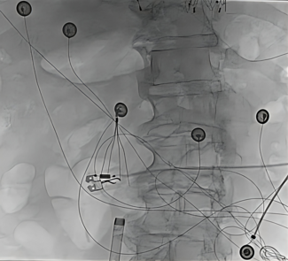
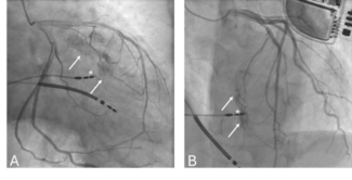
Despite the use of multiple guidewires, including a PT Graphix Intermediate 0.014 (Boston Scientific), a Miracle 3 Straight 0.014 (Asahi Intecc Co. Ltd, Aichi, Japan), through a Maverick (1.5/15 mm) Over-The-Wire balloon (Boston Scientific), it proved impossible to cross the lesion. When the Miracle wire appeared to have taken a subintimal course, a second wire, the Safe-Cross Straight (Intraluminal™ Therapeutics) was advanced parallel to the first (Figure 2). At this point, the Safe-Cross Straight wire was advanced through the over-the-wire balloon to the level of the occlusion, with eventual success. The system was activated and the wire was used to burn a channel through the first few millimeters of the occlusion. Subsequently, this wire was removed and replaced by a PT Graphix Intermediate that easily crossed into the distal vessel.
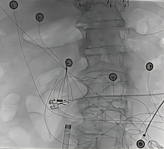
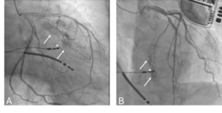
Despite several attempts, the occlusion could not be crossed with any available balloon: Maverick (1.5/15) Over-the-Wire balloon, Worldpass rapid-exchange balloon 1.5 x 30 mm (Cordis Corporation), or Hayate balloon Pro 1.5 x 20 mm (Terumo, Tokyo, Japan). Furthermore, the guiding catheter position was lost. This required a difficult reintubation of the guiding catheter in the presence of a stent protruding into the aorta, and the wire was readvanced across the occlusion; however, the balloon could not be advanced into the ostium. It became apparent that the wire had passed through the struts of the stent that were protruding into the aorta. Thus, a second wire was advanced into the artery and the balloon was advanced without difficulty over this wire to the lesion. However, crossing still proved impossible. As a last resort, we decide to advance a Rotablator wire (Boston Scientific) parallel to the first wire; this did not cross the lesion until the first wire was removed. Then, the Rotawire floppy 0.009 inch wire was successfully manipulated into the distal vessel. A 1.5 mm burr was advanced and easily crossed the lesion (Figure 3). Successive inflations with a Hayate Pro 1.5 x 20 mm and a Maverick 2.5 x 20 mm were followed by placement of a Taxus stent (2.5 x 20 mm) in segment 2. The residual diameter stenosis was 10%, the minimal lumen diameter 1.93 mm, and the reference diameter 2.14 mm, with TIMI 3 flow and a normal blush (Figure 4). The patient was discharged 24 hours later and has since been symptom-free.
Discussion. This case illustrates many of the problems that can be encountered during attempted PCI of a CTO. Although there were somewhat unfavorable angiographic characteristics (blunt stump morphology, side branch at the level of the CTO, and bridging collaterals), the RCA location in conjunction with the absence of other significant lesions, the short length of the occlusion, and its relatively short presumed duration, led us, after consultation with our surgical colleagues, to attempt PCI.
The initial problem was related to the ability to obtain coaxial intubation and adequate guiding catheter support that required the use of multiple catheters in conjunction with implantation of a stent at the ostium. The second problem was the inability to cross the lesion with a wire. Many types of wires are available, and the initial choice is generally a matter of operator preference. Usually, softer tip wires are used first, followed by progressively stiffer wires in order to minimize the risk of perforation.9 When a wire appears to have taken a subintimal course, as occurred in this case, it is sometimes useful to leave it in place and to advance a second wire, in parallel, to find an alternative route.
The Intraluminal™ guidewire is a recently developed technology that delivers radiofrequency energy pulses capable of ablating tissue. In addition, the system monitors, in real-time, the position of the wire of the 0.014 inch wire using optical coherence reflectometry. Near-infrared light is emitted, and by analyzing the amount that is reflected from tissue interfaces, the system is able to determine whether the position of the distal tip of the wire is correct and intraluminal, or too close to the outer vessel wall. Ablation pulses are only allowed when the wire is in the true lumen. Initial studies have demonstrated the utility of this technique to cross CTOs.11,12
The next problem we encountered was the inability to cross the lesion with several different balloons. Attempted balloon crossing resulted in loss of the guiding catheter position, which was resolved with difficulty due to the fact that the stent in the ostium protruded into the aorta. The use of rotational atherectomy finally allowed a balloon to cross and to dilate the lesion. The procedure was completed with the placement of a paclitaxel-eluting stent, our default strategy.
Although the use of such complex technologies undoubtedly increased the procedural costs, such a successful outcome reduces the likelihood that the patient will undergo bypass surgery and improves his long-term survival.2,5 Stenting CTO lesions has been shown to improve long-term outcome; the use of a drug-eluting stent in this diabetic patient with a small diameter vessel reflects current best medical practice.16–22 Preliminary studies suggest that drug-eluting stents remain patent in more than 95% of cases.21
1. Kahn JK. Angiographic suitability for catheter revascularization of total coronary occlusions in patients from a community hospital setting. Am Heart J 1993;126:561–564.
2. Suero JA, Marso SP, Jones PG, et al. Procedural outcomes and long-term survival among patients undergoing percutaneous coronary intervention of a chronic total occlusion in native coronary arteries: A 20-year experience. J Am Coll Cardiol 2001;38:409–414.
3. Ivanhoe RJ, Weintraub WS, Douglas JS, Jr, et al. Percutaneous transluminal coronary angioplasty of chronic total occlusions. Primary success, restenosis, and long-term clinical follow-up. Circulation 1992;85:106–115.
4. Olivari Z, Rubartelli P, Piscione F, et al. on behalf of the TOAST-GISE Investigators. Immediate results and one-year clinical outcome after percutaneous coronary interventions in chronic total occlusions: Data from a multicenter, prospective, observational study (TOAST-GISE). J Am Coll Cardiol 2003;41;1672–1678.
5. Warren RJ, Black AJ, Valentine PA, et al. Coronary angioplasty for chronic total occlusion reduces the need for subsequent coronary bypass surgery. Am Heart J 1990;120:270–274.
6. Maiello L, Colombo A, Gianossi R, et al. Coronary angioplasty of chronic occlusions: Factors of procedural success. Am Heart J 1992;124:581–584.
7. Puma JA, Sketch MH, Tcheng JE, et al. Percutaneous revascularization of chronic coronary occlusions: An overview. J Am Coll Cardiol 1995;26:1–11.
8. Noguchi T, Miyazaki S, Morii I, et al. Percutaneous transluminal coronary angioplasty of chronic total occlusions. Determinants of primary success and long-term clinical outcome. Cathet Cardiovasc Intervent 2000;49:258–264
9. Lefèvre T, Louvard Y, Loubeyre C, et al. A randomized study comparing two guidewire strategies for angioplasty of chronic total coronary occlusion. Am J Cardiol 2000;85:1144–1447.
10. Saito S, Tanaka S, Hiroe Y, et al. Angioplasty for chronic total occlusion by using tapered-tip guidewires. Cathet Cardiovasc Diagn 2003:59:305–311.
11. Chen WH, Lee PY, et al. Initial experience and safety in the treatment of chronic total coronary occlusions with a new optical coherent reflectometry-guided radiofrequency ablation guidewire. Am J Cardiol 2003;15:732–734.
12. Chen WH, Ng W, Lee PY, Lau CP. Recanalization of chronic and long occlusive in-stent restenosis using optical coherence reflectometry-guided radiofrequency ablation guidewire. Catheter Cardiovasc Intervent 2003;59:223–229.
13. Orlic, D, Chieffo A, Stankovic G, et al. Prelimanary experience with the front runner coronary catheter, novel device dedicated to mechanical revascularization of chronic total occlusions. Supplement to J Am Coll Cardiol 2004;43:56A.
14. Gruberg L, Mehran R, Dangas G, et al. Effect of plaque debulking and stenting on short- and long-term outcomes after revascularization of chronic total occlusions. J Am Coll Cardiol 2000;35:151–156.
15. Kiesz RS, Rozek MM, Mego DM, et al. Acute directional coronary atherectomy prior to stenting in complex coronary lesions: ADAPTS Study. Cathet Cardiovasc Diagn 1998;45:105–112.
16. Rubartelli P, Verna E, Niccoli L, et al. for the GISSOC Investigators. Coronary stent implantation is superior to balloon angioplasty for chronic coronary occlusions: Six-year clinical follow-up of the GISSOC Trial. J Am Coll Cardiol 2003;41:1488–1492.
17. Lotan C, Rozenman Y, Hendler A, et al. Stents in Total Occlusion for restenosis Prevention. The multicentre randomized STOP study. Eur Heart J 2000;21:1960–1966.
18. Lemos PA, Serruys PW, van Domburg RT, et al. Unrestricted utilization of sirolimus-eluting stents compared with conventional bare stent implantation in the "real world": The Rapamycin-Eluting Stent Evaluated at Rotterdam Cardiology Hospital (RESEARCH) registry. Circulation 2004;109:140–142.
19. Moses JW, Leon MB, Popma JJ, et al. for the SIRIUS Investigators. Sirolimus-eluting stents versus standard stents in patients with stenosis in a native coronary artery. N Engl J Med 2003;349:1307–1309.
20. Stone GW, Ellis SG, Cox DA, et al., TAXUS-IV Investigators. A polymer-based, paclitaxel-eluting stent in patients with coronary artery disease. N Engl J Med 2004;350:211–212.
21. Hoye A, Tanabe K, Lemos PA, et al. Significant reduction in restenosis following the use of sirolimus-eluting stents in the treatment of chronic total occlusions. J Am Coll Cardiol 2004;43:1954–1958.
22. Nakamura S, Selvan TS, Bae JH, et al. Impact of sirolimus-eluting stent on the outcome of patients with chronic total occlusions: Multicenter registry in Asia. J Am Coll Cardiol 2004;43:35A.


