Clinical Images
Identical Twins, Identical Coronary Disease
December 2005
Background. A number of studies have examined the environmental and genetic basis contributing to the pathogenesis of various disease states. This has been studied in monozygotic twins and recently published. Recent reports have examined disease prevalence, mechanism of onset, and disease progression in large cohorts of twins as it pertains to insulin resistance states,1 congenital heart disease,2 Parkinson’s and other neurologic disease states,3 as well as gastroesophageal reflux disease,4 to name just a few. Although endocrine disease is thought to generally fit a multifactorial pattern of transmission, twin studies have clearly established a higher degree of genetic susceptibility.5–8 Despite this, there remains considerable controversy regarding the relative contribution of environmental factors versus genetic predisposition in a variety of disease states.
The incidence of coronary disease in identical twins has not been clearly established. A review of the existing literature describes a largely anecdotal experience, published primarily in the form of case reports over the last decade.9 Though large-scale epidemiologic twin studies are lacking and have not clearly established a link in cardiovascular disease, recently published case reports have been compelling.10
We report a case of identical twins presenting with unstable angina within a week of each other, and in whom cardiac catheterization demonstrated nearly identical coronary artery lesions. Moreover, their pattern of angina and symptom onset at acute presentation were also strikingly similar.
Clinical Presentation. A 45-year-old male presented with symptoms of chest tightness. He had no documented history of diabetes, hypertension or hyperlipidemia. The patient was a nonsmoker and there was no documented family history of premature coronary disease.
Previously, he had a single episode of atypical, right-sided chest discomfort and underwent outpatient noninvasive testing. Two previous outpatient stress tests had been performed, presumably initially for what was described as “atypical chest symptoms”. The first study was reported as being normal following 10 minutes of exercise according to the Bruce Protocol. Myocardial perfusion imaging showed a homogeneous perfusion pattern and no obvious areas of ischemia. However, four weeks later a second study, performed again due to “atypical symptoms”, suggested septal ischemia on perfusion imaging. Once again, an exercise duration of 10 minutes was achieved with no anginal symptoms reported. The patient declined any further invasive studies. No specific therapy was initiated following the second stress test, with the exception of statin agents for hyperlipidemia, as well as low-dose beta blockade for mild hypertension.
Approximately four weeks later, the patient presented acutely with new-onset rest symptoms of angina. The symptoms were described as chest tightness with radiation to the neck and arm in contradistinction to the atypical symptoms noted at the time of initial outpatient evaluation. Mild dyspnea was noted in association with chest discomfort. He was triaged to diagnostic coronary arteriography.
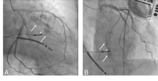
Cardiac catheterization, shown in Figure 1A, demonstrated high-grade tandem lesions in the left anterior descending coronary artery (LAD). A 3.0 x 28 mm drug-eluting stent was implanted in the LAD without complication. The postintervention images are seen in Figure 1B. The patient was subsequently discharged, with the addition of combination antiplatelet therapy to the existing regimen.
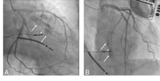
Approximately one week later, the identical twin brother of the patient described above presented with similar symptoms of rest angina. An avid basketball player, he had noticed mild “right-sided” symptoms with exertion several weeks earlier. However, this was his first episode of “chest tightness” with minimal activity. Symptoms were associated with radiation to the neck and mild dyspnea. Cardiac catheterization was performed (Figure 2A), showing tandem high-grade LAD lesions. Deployment of a 4.0 x 33 mm stent at the lesion site was performed, with further stent dilatation using a 4.5 x 15 mm noncompliant balloon (Figure 2B). The patient was discharged on a statin agent, beta blockers and combination antiplatelet therapy. At outpatient follow-up, both patients were stable and remained free of symptoms.
Discussion. The exact mechanism for the development of coronary disease in identical twins has not been clearly defined. Certainly, the constellation of clinical events described above, as well as the associated clinical descriptors ought to be considered more than merely coincidental and provides a provocative case for the genetic transmission of coronary artery disease.
Several recent publications have described potential genes responsible for the transmission of coronary disease.11–13 It has been postulated that multiple distinct genetic complexes may be responsible for the transmission of atherosclerosis. These include “causative” genes, as well as those described as “disease-susceptibility” genes. Unlike Mendelian genetics, in which phenotypic expression is the result of a more simplified interplay of dominant or recessive genes, cardiac disease states may represent a significantly more complex association of disease modifying genes with specific environmental influences incriminated as risk factors.11 Equally compelling, under optimal clinical circumstances, this theoretical interdependence of genetics and environment may also confer protection from certain disease states as well.
Kaluza et al.10 have previously described a similar case involving identical twins who demonstrated nearly identical LAD disease that necessitated percutaneous coronary intervention. However, in that case report, the interval between clinical presentations of each twin patient was separated by a longer period of time. Our case is even more provocative because the clinical timing, presentation, symptoms, coronary anatomy, culprit lesions and even their presenting ECGs (Figures 3A and B), were all nearly identical. Interestingly, Kaluza makes the point that the strong similarity in the coronary anatomy may have in some way “fated” the ultimate location of lesion formation.10
Similar to previous case reports (which comprise most of the available literature), the onset of symptoms in our identical twin patients was at a relatively young age, there was striking similarity in baseline electrocardiography (Figures 3A and B), and the findings at the time of coronary angiography were similar. This triad of clinical descriptors is consistent with the observations of previous authors.9 It should also be mentioned that the described electrocardiographic similarities may be more striking in the anterior precordial leads.
Nathoe et al. recently published an analysis that comprehensively reviews the currently available studies (and case reports) on twin concordance and the development of symptomatic atherosclerosis.14 This retrospective analysis suggests that there is a strong similarity amongst identical twins who have developed coronary disease relative to the age of onset of clinical manifestations. It also seems to reinforce the concept of “early age” at the time of initial diagnosis. In those studies and case reports that were reviewed, the average age of the first coronary event was 42 ± 12 years. Other recognized similarities in these twin studies include coronary risk factor profile as well as the “type of coronary event” at the time of initial clinical presentation.
Conclusion
The study of cardiovascular disease patterns in identical twins should be more than simply a case of clinical fascination. More comprehensive and larger-scale studies may help to unlock some of the mysteries of the genetic basis for cardiovascular disease and assist in our ever-increasing need for adequate patient counseling and primary prevention.
1. Rasmussen F, Johansson-Kark M. The Swedish young male twins register: A resource for studying risk factors for cardiovascular disease and insulin resistance. Twin Res 2002;5:433–435.
2. Zaghloul N, Hutcheon RG, Tepperberg JH. Visual diagnosis: Monozygotic twins who have congenital heart disease and distinctive facial features. J Intern Med 2002;252:247–254.
3. Tanner CM, Goldman SM, Aston DA, et al. Smoking and Parkinson’s disease in twins. Neurology 2002;59:1821–1822.
4. Cameron AJ, Lagergren J, Henriksson C, et al. Gastroesophageal reflux disease in monozygotic and dizygotic twins. Gastroenterology 2002;122:55–59.
5. Brix TH, Christensen K, Holm NV, et al. A population based Study of Graves’ disease in Danish twins. Clin Endocrinol 1998;48:393–395.
6. Tani J, Yoshida K, Fukazawa H, et al. Hyperthyroid Graves’ disease and primary hypothyroidism caused by TSH receptor antibodies in monozygotic twins: Case reports. Endocr J 1998;45:117–121.
7. Dubrey SW, Reavely DR, Seed M, et al. Risk factors for cardiovascular disease in IDDM. A study of identical twins. Diabetes 1994;43:831–835.
8. Kumar D, Gemayel NS, Deapen D, et al. North American twins with IDDM. Genetic, etiological, and clinical significance of disease concordance according to age, zygosity, and the interval after diagnosis in first twin. Diabetes 1993;42:1351–1363.
9. Samuels LE, Samuels FS, Thomas MP, et al. Coronary artery disease in identical twins. Ann Thorac Surg 1999;68:594–600.
10. Kaluza G, Abukhalil JM, Raizner AE. Identical atherosclerotic lesions in identical twins. Circulation 2000;101:E63–E64.
11. Kraus WE. Genetic Approaches for the investigation of genes associated with coronary heart disease. Am Heart J 2000;140:S27–S35.
12. Winkelmann BR, Hager J, Kraus WE. Genetics of coronary heart disease: Current knowledge and research principles. Am Heart J 2000;140:S11–S26.
13. Hauser ER, Mooser V, Crossman DC, et al. Design of the genetics of early onset cardiovascular disease (GENECARD) study. Am Heart J 2003;145:602–613.
14. Nathoe HM, Stella PR, Eefting FD, et al. Angiographic findings in monozygotic twins with coronary artery disease. Am J Cardiol 2002;89:1006–1009.


