Case Report
Coronary Fistula from Left Main Stem to Main Pulmonary Artery
December 2003
Congenital coronary arteriovenous fistula (CAF) is a rare anomaly. The incidence of congenital cardiac lesions is only 0.13%.1 Over 90% of these fistulas drain into the systemic venous side of the circulation.2 Drainage of the fistula into the pulmonary trunk has been reported in 17% of cases.2 To the best of our knowledge, a connection between the left main stem and main pulmonary artery has been reported in the literature only once, as a case in India in 1989.3
Case Report. A 51-year-old female with hypertension was admitted to the hospital with her first attack of acute myocardial infarction (AMI). There was no evidence of heart failure. The electrocardiogram revealed sinus rhythm with Q-waves on precordial leads V2 to V4, with a negative T-wave. Maximal creatine kinase elevation was 1,500 IU at 24 hours after the onset of clinical symptoms. During the hospitalization, the patient had 2 episodes of post-infarction angina.
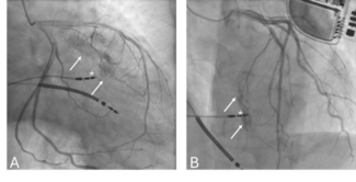
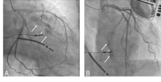
Coronary arteriography showed a significant 90% obstruction in the middle segment of the left anterior descending (LAD) coronary artery and diffuse atherosclerosis in the other vessels. Percutaneous transluminal coronary angioplasty with the implantation of 1 stent type (BESTENT-2) was successfully performed. Normal flow was restored. Coronary arteriography revealed a fistula connection arising from the left main stem and draining into the main pulmonary artery (Figure 1).
Discussion. Coronary arteriovenous fistula (CAF) arising from the left main artery is a very uncommon entity. In a review of 363 cases with coronary arteriovenous fistulas,2 fifty percent were found to arise from the right coronary artery, forty-two percent from the left coronary artery and 5% from both coronary arteries. Origin from the left main coronary artery was rare.4 Approximately 5% of coronary arteriovenous fistulas are bilateral.2 The most common site of drainage is the right ventricle (41%), followed by the right atrium (26%) and pulmonary artery (17%).2 When bilateral, fifty-six percent of these fistulas have been found to drain into the pulmonary artery.5 However, only 1 case of a fistula connecting the left main coronary artery (LMCA) and pulmonary artery has been previously reported,3 although other reports connecting the LMCA to right heart chambers are available.4,6 Patients with CAF may have symptoms of pulmonary congestion due to left to right shunt or they may be asymptomatic. Complications, such as congestive heart failure, infective endocarditis and myocardial infarction, also have been reported.2,7
Management of CAF is controversial, particularly in patients with small shunts. Liberthson7 reviewed 187 cases and found that operative mortality (1%) and complications (7%) were low if patients were operated upon at the age of 20 or younger. However, in those who were operated upon after the age of 20, significant post-operative mortality (7%), post-operative myocardial infarction (7%) and other complications (23%) occurred. The authors recommended surgery in all cases of CAF in childhood, irrespective of symptoms or size of the shunt.8–11
A new therapeutic option for the treatment of CAF is the percutaneous transcatheter embolic occlusion or transcatheter closure (TCC) technique, which has been advocated as a minimally invasive alternative to surgery. A variety of materials have been used, including Gianturco coils, covered stainless-steel coils, detachable balloons, coaxial embolization with platinum microcoils, double-umbrella devices and the Gianturco Grifka vascular occlusion device.8,12–14
Feasibility of closure by a device is determined by the number and location of drainage sites, the ability to cannulate the distal fistula and the proximity of coronary branches to the optimal occlusion site. Special care should be taken to avoid allowing the device to interfere with flow into any visible coronary vessel. The selection of occlusion device was based upon the anatomic features of the fistula.11 Coils have been used in small CAF, while double-umbrella devices have been used in larger CAF. Device deployment has been performed either anterogradely (via the femoral vein) or retrogradely (via the femoral artery).15 Based on the TCC literature and on their own data, Armsby et al.12 suggested that 40% of patients with larger CAF were symptomatic (dyspnea on exertion, angina, congestive heart failure, fatigue and palpitations). A continuous murmur was heard in most of these patients.
Successful occlusion of CAF at catheterization was reported in 83% of patients. Procedural complications included a similar number of instances of transient ischemic changes on the electrocardiogram and unretrieved device embolization to the tricuspid valve or distal pulmonary artery, and a single incidence of myocardial infarction and transient atrial arrhythmia. There were 2 procedure-related deaths (2.2%).
Follow-up data is available for 42 of 45 patients in the TCC literature12–17 (mean follow-up, 12 months). All were asymptomatic and there were no late complications or deaths. Follow-up imaging studies (echocardiogram in 27 patients, angiogram in 6 patients) showed complete closure in 91% of patients.
Some features that may render CAF patients unsuitable for TCC include extreme vessel tortuosity, multiple drainage sites and coronary branches at the location of optimal device deployment.12 The ability to cannulate the distal fistula and avoid flow interference through nearby coronary branches is mandatory for successful TCC.12 Armsby et al.12 have adapted the following therapeutic strategy for CAF patients: 1) those with CAF and additional complex heart disease requiring surgery are referred for surgical repair; 2) patients with clinically significant CAF are referred for catheterization, where the fistula anatomy is further defined; and 3) those with suitable anatomy undergo TCC, while those who are unsuitable are offered surgical ligation or are medically followed.
Conclusion. With increased experience and improved devices and techniques, TCC of CAF is emerging as a successful therapeutic strategy. The preferred approach for any individual patient will depend on the anatomy of the fistula, the presence or absence of associated defects, and the experience of the interventional cardiologists and surgeons.
1. Gillibert C, Van Hoof R, Van de Werf F, et al. Coronary artery fistulas in an adult population. Eur Heart J 1986;7:437–443.
2. Leven DC, Fellow KE, Abrams HL. Hemodynamically significant primary anomalies of the coronary artery. Circulation 1978;58:23–34.
3. Sethi KK, Prasad GS, Arora R, Khalilullah M. Coronary arterio-venous fistula from left main stem to pulmonary artery. Indian Heart J 1989;41:194–195.
4. De Nef JJE, Varghese PJ, Losekoot G. Congenital coronary artery fistula — Analysis of 17 cases. Br Heart J 1971;33:857–861.
5. Fujiwara R, Kutsumi Y, Yamamura I, et al. Bilateral coronary arteriovenous fistulas associated with idiopathic hypertrophic cardiomyopathy. Am Heart J 1986;111:1207–1211.
6. Gupta SK, Abraham KA, Moorthy JSN, et al. Left main coronary artery-right atrial fistula. Indian Heart J 1985;37:308–309.
7. Liberthson RR, Sagar K, Berkoben JP, et al. Congenital coronary arteriovenous fistula. Circulation 1979;59:849–854.
8. Mavroudis C, Backer CL, Rocchini AP, et al. Coronary artery fistulas in infants and children: A surgical review and discussion of coil embolization. Ann Thorac Surg 1997;63:1235–1242.
9. Carrel T, Tkebuchava T, Jenni R, et al. Congenital coronary fistulas in children and adults: Diagnosis, surgical technique and results. Cardiology 1996;87:325–330.
10. Goto Y, Abe T, Sekine S, et al. Surgical treatment of the coronary artery to pulmonary artery fistulas in adults. Cardiology 1998;89:252–256.
11. Schumacher G, Goithmaier A, Lorenz HP, et al. Congenital coronary artery fistula in infancy and childhood: Diagnostic and therapeutic aspects. Thorac Cardiovasc Surg 1997;45:287–294.
12. Armby LR, Keane JF, Sherwood MC, et al. Management of coronary artery fistulae. Patient selection and results of transcatheter closure. J Am Coll Cardiol 2002;39:1742–1744.
13. Dorros G, Thora V, Ramidreddy K, Joseph C. Catheter-based techniques for closure of coronary fistulae. Cathet Cardiovasc Intervent 1999;46:143–150.
14. Reidy JF, Anjos RT, Qureshi SA, et al. Transcatheter embolization in the treatment of coronary artery fistulas. J Am Coll Cardiol 1991;18:187–192.
15. Pedra CAC, Pihkala J, Nykanen DG, Benson LN. Antegrade transcatheter closure of coronary artery fistulae using vascular occlusion devices. Heart 2000;83:94–96.
16. Harris W, Andrews J, Nichols D, Holmes D. Percutaneous transcatheter embolization of coronary arteriovenous fistulas. Mayo Clin Proc 1996;71:37–42.
17. Qureshi SA, Reidy JF, Alwi MB, et al. Use of interlocking detachable coils in embolization of coronary arteriovenous fistulas. Am J Cardiol 1996;78:110–113.


