More Insights on the Convergent Talus
Key Takeaways
Convergent Talus Is a Critical Driver of Complex Foot Deformity Across Multiple Pathologies
The podcast emphasizes that convergent talus is not a diagnosis, but a pathomechanical pattern seen in conditions such as talar neck malunion, Charcot midfoot collapse, perinavicular pathology, and neuromuscular disease. When the talar neck shortens or medial column length is lost, the foot shifts into a closed-chain supinatory state, resulting in heel varus, forefoot adduction, and abnormal load transfer to the lateral column. Recognizing talar convergence early allows clinicians to better predict deformity progression and avoid delayed or incomplete reconstructions.
Surgical Planning Must Address Medial Column Length and Joint Mobility—Not Just Alignment
Dr. Visser highlights that failure to restore talar neck length or mobilize locked joints leads to persistent deformity even after corrective procedures. In talar neck malunion, medial column shortening often requires interpositional bone grafting, while fixed subtalar or talonavicular joints may necessitate capsular releases, osteotomies, or triple arthrodesis. Similarly, in Charcot foot, talar position relative to the calcaneus and midfoot determines whether conservative care is appropriate or whether early reconstruction is required to prevent rocker-bottom ulceration.
Loss of Peroneal Function Creates a Predictable Convergent Talus Deformity Requiring Active Tendon Transfer
When both peroneal tendons fail, the resulting adductovarus foot and convergent talus cannot be corrected with allograft alone if muscle function is compromised. The discussion outlines how FDL tendon transfer, combined with heel varus correction (e.g., Dwyer osteotomy) and selective tibialis posterior weakening, restores balance by reintroducing an active lateral motor. Intraoperative muscle stimulation and preoperative MRI assessment are essential to determine whether a functional transfer—or a salvage procedure—is indicated.
Transcript
Please note: This content is a direct transcript, capturing the authentic conversation without edits. Some language may reflect the flow of live discussion rather than polished text.
Jennifer Spector, DPM: Welcome back, everybody, to Podiatry Today Podcast, where we bring you the latest in foot and ankle medicine and surgery from leaders in the field. I'm Dr. Jennifer Spector, the assistant editorial director for Podiatry Today. And with this episode, we're continuing our series with Dr. Visser on the convergent talus. We've touched on many podiatric conditions that really fall under this umbrella and each one has very unique considerations for us to keep in mind. So we'll continue the conversation today with Dr. Visser in the same vein. And just to reintroduce you to his expertise, he is a past president of the Missouri Podiatric Medical Association and the St. Louis Podiatric Medical Society. He has served as an examiner for the American Board of Podiatric Surgery and on the Missouri State Board of Podiatric Medicine for several years. He is a long-term residency director with hundreds of residents that he is trained and was inducted in 2015 into the Kent State University School of Podiatric Medicine Hall of Fame.
So without further ado, let's dive into this next consideration with the convergent talus. So what about malunion of the talar neck? Can you explain how that factors into all of this?
H. John Visser, DPM: Malunion of the tailor neck is something that you hear a lot about, but you don't really see much about how do you diagnose it? How do you actually treat it? You know it's a potential complication that occurs, but you really don't understand what's actually happening there. Obviously, we have plenty of lectures on talar neck fractures and how they're approached and how they're treated. But I can't really even think of anybody that really talks about a malunion. What happens in a malunion? Well, normally, as we know, the mechanism of fracture of the talar neck is usually from a dorsiflexion inversion mechanism. So what happens is, in that particular instance, comminution tends to occur on the medial side of the talus, where the lateral side, the fracture is a complete fresh linear fracture. So that's the reason when you do talar neck fractures, you always approach the lateral side of the neck first to basically realign it. And by realigning the lateral side of the talar neck, you have it in anatomic alignment.
You then go to the medial side, which oftentimes will be comminuted and then address the comminution with grafting and then use a plate to basically realign the neck in its corrected alignment. What happens in the case of a malunion, the medial side of the fracture is not really properly addressed. In other words, they may just go in and percutaneously run two screws and try to compress the fracture. And when they do, they cause shortening of the talar neck. Now, what happens when the talar neck is short is it will create a closed kinetic chain supinatory effect. And also what will happen is the forefoot will go into a varus attitude and it will be adducted. So we'll have an adducted and we'll have a varus talonavicular joint, and we will have close kinetic chain supination and convergence of the talus.
Once that heals, the patient begins a year or two afterwards to function with a fixed heel varus and they begin to walk on the lateral side of their foot. At times, the medial side of the foot is not really weightbearing or you notice a functionally plantarflexed first metatarsal created by the fact that the peroneus longus now has this big mechanical effect to pull the medial side of the foot to the ground. The medial side of the foot always wants to get to the ground no matter what.
So this is the situation you have, and it makes it a very difficult problem to actually correct because once you have this problem, you have to go to the medial side in an attempt to lengthen it. And this usually requires an interpositional bone graft, usually an allograft or an autograft to lengthen this.
Now, also, when the lengthening process occurs, the subtalar joint will go through a closed kinetic chain pronatory effect, but sometimes it doesn't. Sometimes it's stuck there. And as you lengthen the neck, it stays fixed in that position. Also, similarly, the talonavicular joint may stay in that fixed position where it is in a varus attitude.
Jennifer Spector, DPM: What happens in those cases?
H. John Visser, DPM: In that case, you're basically relegated to a triple arthrodesis because you have to realign the talonavicular joint, you have to realign the subtalar joint, and in that case, that's going to require a form of a triple arthrodesis. There are some instances though with the lengthening process and the way you do, you lengthen it with a laminar spreader or a Hintermann retractor, and then you see what you've got. Is the heel varus corrected at this particular alignment? If it's not, you can then attempt at the subtalar joint level to basically, since you have the medial side open at that point with your incision, to do a release of the capsule of the subtalar joint and an attempt to mobilize it in that particular instance. And in some instances, you can release it and the heel varus can be corrected or you're getting some mobility. Maybe it doesn't fully correct, but you're getting some mobility.
And if it's not fully corrected, but you get the mobility, then a Dwyer osteotomy can be effective. Similarly, at the talonavicular joint, you have to go in and do a complete pancapsulotomy of the talonavicular joint in that particular situation to get to mobilize the talonavicular joint to move from this adducted and varus state. And it usually requires, if you are able to reduce this, you got to abduct it and you got to de-rotate it. You usually need to place a Steinmann pin in for a period of at least about four weeks so that the soft tissues heal into its corrected position. So you can see it's a really difficult problem to deal with if you have it.
And again, you've heard about, "Oh yeah, that's a complication. Always run it through." But nobody ever explains, well, what do you do if you have the problem? What can you possibly do in that particular situation? And that's the mechanics behind it. Yes, the talus is, again, convergent created by shortening of the medial column of the talus. The talus, the neck is short and this creates a closed kinetic chain supinatory effect. But because the pathology here, the pathomechanics here, the old adage, well, the subtalar joint, the calcaneal cuboid joint moves with the subtalar joint, that is true. But here, the talonavicular joint, which should go into a more of a supinatory state basically goes into a varus type of an attitude, and then obviously the procedure needs to be addressed by elongating what was lost in comminution.
Jennifer Spector, DPM: What about Charcot foot? I'm sensing a theme here about when this may come into play, and I'd be curious to know your thoughts on where that falls into the scope.
H. John Visser, DPM: The Charcot foot is very interesting, and I'm talking about the midfoot form, and the midfoot form obviously is the most common. And as we know, with the midfoot condition, now the idea in the case of a stage one instance is the idea is we should go ahead and fix those. We should treat them like a Lisfranc's dislocation as opposed to the old adage where you would go ahead and sit on those until they got into a Eichenholtz stage 3 and then determine what the foot position is, where is it? Is it going to create an ulcer? Can I get it in a shoe? Then do I need to do a reconstruction?
Very interesting thing that I have observed over time that I look for to determine whether or not I think this particular foot probably is going to need surgery or it may not. You also are aware of many of the midfoot types of Charcot that they go through their process, they go through stage 1, stage 2, stage 3 of a Eichenholtz, and they end up with basically a foot that's plantargrade and they don't really have any excessive weightbearing points that become ulcerative. They don't have a rocker bottom situation. And because they've been able to maintain the forefoot to rear foot alignment, they're able to be shot in usually extra depth shoes. So there are those people.
And again, unfortunately, I will get people referred to me who, because of the way their x-rays look, that they need to have a Charcot reconstruction. Yet you look at them clinically, they don't have a rocker bottom. They do not have any significant prominence to the plantar surfaces of their foot that would be potentially weightbearing. And they have enough of a forefoot–rearfoot alignment that is in line that they could easily fit in an extra depth shoe. I'm sure you've seen many of these feet, and you're not going to do a surgical reconstruction on them because of the way the foot looks. Yeah, it looks terrible, but you're not going to do a reconstruction because you got to plant a grade foot and you're basically able to fit them in traditional types or in extra depth types of shoewear and sometimes even normal shoe wear.
Jennifer Spector, DPM: Can you tell me more about some unique observations when it comes to the stage 1 Charcot patients here?
H. John Visser, DPM: Where is that talus? Where is the talus to the calcaneus? Now, in instances where it's divergent and the talus is abducted, if that particular case ensues, and I see that, I'm going to want to fix that because I know down the road I'm going to get a rocker bottom, or I might try an Achilles or a gastroc early on to unload the midfoot.
If I see, on the other hand, the talus basically centrally located aligning with TMT 2–3, that foot is going to end up with a plantargrade foot. It's going to be a plantargrade foot. Because of its alignment with the central column, the central column does not become into a recurvatum state. And as its cuneiforms undergo avascular necrosis and go through that process as they go in from a Eichenholtz stage 2 and 3. And that particular instance, I'm not going to do anything with that. I won't even do the primary procedure because I know that that foot's going to end up plantargrade. Then I'll get one that has a convergent talus, usually due to a perinavicular issue. We know the perinavicular issues can occur in Charcot and are very similar to the discussion we talked about with Charcot-Marie-Tooth disease, although they also have a lot of cuneiform disease and so forth involved with them. And in that case, with the talus being convergent and over on the cuboid fourth and fifth articulation, I'm going to end up with a cuboid rocker bottom. And we all know when we have the cuboid prominent on the plantar aspect of the foot, there's nothing you can do for that other than to operate on it because it's going to end up with an ulceration. Equinus releases don't help that, and I'm going to go ahead and I'm going to operate on that.
So that's an observation I have come across over my years of looking at Charcot feet and kind of give me an idea. Am I going to get myself involved in this in stage 1, and do I need to, or can I sit this one out? And that's kind of what I do when I look at Charcot. So convergence of the talus there is very important as is divergence in this particular instance.
Jennifer Spector, DPM: To round out this list of conditions that really play into this theme of the convergent talus, what happens when, whether it's due to traumatic reasons or neuromuscular reasons, what happens then when you lose that peroneal tendon function?
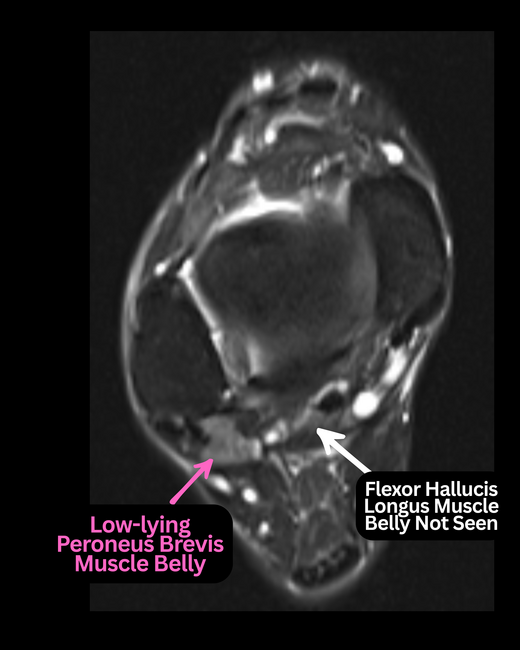
H. John Visser, DPM: When you have peroneal tendinopathy where both peroneal tendons are involved with tendinosis, and how can this happen? It's a relatively rare condition, but it can happen. Most of the time it's usually from failures of index operations. The patient has an operative procedure, usually of the brevis, and they may have the lungs done at that time, and the patient has a failure of that particular condition. And basically you end up with a patient has significant thickening over on the lateral side of the ankle on the fibula. You get an MRI on it, and oftentimes it will show a tear or rupture of the brevis at the retrofibular groove. And oftentimes the peroneus longus too will show a rupture in the area of the cuboid groove. And as a result of that, you basically have a zero cable effect. You have no peroneal tendons on that side of the foot.
Now, the thing is, do you have muscle function? One of the ways you can determine it preoperatively is to get MRI in the area of the peroneal musculature and look at things such as fibrosis or fatty infiltration. If you begin to see changes like that, it indicates that the muscular group itself is not really highly functional. If on the other hand, you got that peroneal or you got the MRI and it did show any of that, and you took a look at the patient clinically and you looked at the girth of their calf and it looked relatively normal on both sides. That case, you could get away with an allograft. You can go ahead, go and excise both of the peroneal tendons, place one allograft. You can use two if you'd like, but one is usually necessary, and you have the full function of the peroneal musculature.
Jennifer Spector, DPM: Are there any ways that you can assess this in the OR?
H. John Visser, DPM: You also intraoperatively want to check that muscle function by basically stimulating it with a monopolar Bovie and see if there's actual contracture of the musculature and palpate the musculature and see if there's any areas of scarring and thickness that would indicate that there's intratendinous scarring going on. If you have these issues where you have fatty infiltration, you have fibrorosis, you can't depend on allograft in that case because the muscles are not working. It's like hitching a dead horse to a wagon, it's not going to pull. What's going to happen here is you're going to have to get an active motor to that side of the foot. And obviously, what type of foot you have here is, again, another form of a convergent talus.
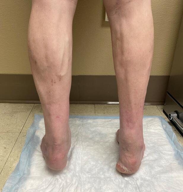
Now, the convergence is usually an adductovarus. It's not a highly adductocavovarus because the peroneals, even though they may not be functional, are still intact and maintaining tension. And as long as you're maintaining tension, there is some resistance to the antagonist, which is obviously, in this case, the tibialis posterior and the tibialis anterior tendon, mainly the tibialis posterior. So you will see a patient who will have adductovarus foot. In other words, the talus will be convergent and you will see a bit of an increase in the calcaneal inclination angle. You'll see a varus heel. In that case, what you have to do is you have to excise obviously the peroneal tendons themselves, they're basically not effective. They can't be repaired and they can't be replaced by allograft. You then take a motor unit on the medial side of the foot and ankle.
Now, as we know, the peroneals are plantar flexors and inverters, and on the medial side, we have the plantarflexors and the inverters. So what are potential options? The tibialis posterior would be one that would be one you would highly think of, but it tends to be relatively short tendon and would not really be able to give you the amount of excursion you need to be transferred over to that side of your foot. So the two options you have are either the flexor digitorum longus, which has the longest tendinous unit or the FHL. Either one—you can use either. Again, the issue with the FHL becomes the neurovascular bundle. It can possibly be pinched off in the transfer. So most people elect to use the FDL tendon. And what you do, then you make an incision at the navicular tuberosity, and you follow the FDL tendon down to the master knot. You then cut it at that level. And the way to give you as much tendon length as you possibly can, you adduct, you plantarflex the foot, curl the toes and give as much as you're pulling on, the FDL will give you about as much tendon as you possibly will need.
You then are able then to make an incision at the ankle level about 9 cm above the ankle joint. And at that point, the first myotendinous tendon you will see will be the FDL. If you're considering when you were talking about the tibialis posterior transfer, you have to be careful about that because the TP tendon is the second tendon down, and that's recruited about usually 12 cm above the ankle, so it's higher up. But you then are able to grab the FDL tendon at that second incision site and pull it through proximately. You then take the peroneals have been opened up. You have an incision on that side of your ankle because you had to excise the entire peroneal tendonous structures, and they have to be excised because they can be definitely pain generators.
Then what you're able to do is you take that FDL tendon and you transfer it from the medial compartment into that lateral compartment. You want to hug the tibia, you want to stay tight up against the tibia. Now, when you made your incision for the FDL transfer and you are going to harvest it in that particular area, you are then able to get access and make a small incision there to the medial side of the tibia. You then pass it into the lateral compartment and then run it underneath the retrofibular groove and take it down to the fifth met base.
Now what I do is I always leave some of the tendon attached to the fifth metatarsal base of the peroneus brevis tendon because that way I can Pulvertaft the FDL transfer and have plenty of length there. If you do get a situation where you end up short on the tendon, you can do what's called a turndown technique where you split the tendon in two halves and then pull it down, reinforce where the tendon basically is cut down to that level, reinforce that, and then it'll give you plenty of tendon in that situation. But if you leave enough of the brevis tendon, you're not going to run into that trouble.
Then you go ahead, and in this case, I'll dorsiflex it and evert it and maximally tension it through a Pulvertaft technique. There's no way that you can overtension that. And in that particular instance, that would be able to complete your transfer. And this is a phasic transfer because you're basically taking plantarflexors and then one's an invert/everter, but the frontal plane doesn't make much of a change or a problem for you. In this case, it's the fact that it's a plantarflexor in this situation. The other thing is too, is to remember the peroneals are one of the muscle tendon units that are also known to undergo phase conversion. That's basically what needs to be done in this situation. You usually have to include a Dwyer osteotomy to correct the heel varus in this particular situation.
And then also it's important that the tibialis posterior tendon be weakened either by a tenotomy, a full tenotomy, or by a Z-plasty lengthening. Remember, any type of a convergent foot is a stiff foot. It's a foot that doesn't move well or evert much as far as at its subthaler joint level. That is something that you should check preoperatively, see how much eversion you have to the subtalar joint. If you do have an instance where it appears that you can evert the subtalar joint easily past vertical, then you should stay away from a tenotomy. You should do a lengthening. But if you can not get it fully everted and you have to do the Dwyer and you're doing the Dwyer, and after the Dwyer, you still can't evert it past vertical, then it's certainly easy to do the tenotomy. Otherwise, that particular tendon unit will potentially create deformity again, and you have to avoid that.
Plantar fascial releases are done dependent on your relative arch height that you have available at that particular time. Now, the antagonist to the peroneus longus, the TA tendon, is usually not that much of a player in this particular instance. Usually you don't have to do anything with the TA as far as the lengthening it or transferring in this particular condition.
Jennifer Spector, DPM: Great. Well, we've gone into quite a few different clinical scenarios where clearly the convergent talar situation is very impactful for everything from surgical planning to managing patient expectations. Is there anything else that you'd like to share on this topic?
H. John Visser, DPM: The only other thing I would probably talk a little bit about, and it dealt with the CMT foot about that non-phasic transfer for a non-phasic transfer to really have the ability to create functional motion of the ankle joint, it has to undergo phase conversion. And by just transferring to the top of the foot and tensioning it at neutral, which is at the ankle at 90 degrees, if you tension it at that level and it doesn't go through a biofeedback mechanism of phase conversion, what will happen is that all that will serve is a tenodesis. It will just basically hold the ankle in that position, and you are not going to get any type of dorsiflexion force out of it. Now, one of the things too with these transfers is they do usually lose about two grades of muscle power when they do it. The big thing about the tibial posterior transfer is the deforming force and the severe deforming force it has on the foot and basically how it drives the muries angle proximally.
So what you have to do is teach the patient what that muscle tendon does. So you can do it either yourself, which I do, or you can send them to a therapist if you like. But what you do is you teach the patient what the tibialis posterior tendon does. We know it's a stance phase muscle, so it doesn't have any real active swing phase muscle power, and it doesn't actually function other than in a stance phase–type of functionality. So we know though what it does do, it's a plantarflexor and inverter, and basically we know it works the retrograde to help externally rotate the tibia to prepare it for propulsion. So what we want to do then is teach the patient what it does, plantarflex and invert. You teach them plantarflex invert, and they pick it up pretty quickly, plantarflex invert.
Then what happens after the transfer's done, and at about two to three weeks when you're going to begin to begin active motion of that muscle tendon unit, you then, and the therapist, if they're not familiar with it, then you actually teach them. What they do is they tell the patient, move that TP muscle, which means plantarflex and invert. So they think plantarflex invert while the therapist dorsiflex and everts. And this process goes on for about two to three weeks and eventually phase conversion occurs that now when they think about plantarflexing and inverting their foot, the foot will actually dorsiflex and abduct, so they will get some dorsiflexion power. Now, certainly most people are going to need to be placed in an AFO type of brace, which is another interesting thing that I'll talk just a little bit about from my resident standpoint.
When I talk about bracing and I say, "Well, if I've done a TP transfer and I want to basically put them in a brace, what type of brace am I going to put them in? Am I going to put them in a total ankle AFO, or am I going to put them in a posterior spring AFO?" A total ankle AFO is where it's extended beyond the malleoli. In other words, the main portion of the AFO extends beyond the malleoli. The posterior springs are posterior to the malleoli, so they have that hinge effect. So obviously if you're going to want the patient to have any type of dorsiflexion, you're going to want to put them in a posterior spring AFO, as opposed to a toll ankle AFO, which you would put, for example, a patient who had a completely osteoarthritic ankle joint, had severe pain in their ankle and you wanted to lock up their motion.
So those are a couple things I think that are important to discussing some of this as far as some adjuncts there. I think the main thing to remember is that how the foot functions and how pathomechanically it will compensate and begin to compensate for pathology in different regions of the foot. We see it at Mueller Weiss, we see it with a talar neck malunion. We certainly see the most complex form, which is Charcot-Marie-Tooth disease. And so I think hopefully this discussion gave you a much better idea about what the term convergent talus actually means and how it can be utilized.
Jennifer Spector, DPM: Well, thank you so much, Dr. Visser, for this really comprehensive series on the convergent talus. You can find this in other Podiatry Today podcasts on our website at podiatrytoday.com, SoundCloud, Spotify, Apple, podcasts, and more. We hope you'll join us next time.
© 2026 HMP Global. All Rights Reserved.
Any views and opinions expressed are those of the author(s) and/or participants and do not necessarily reflect the views, policy, or position of Podiatry Today or HMP Global, their employees, and affiliates.