Video Supplement: Transcatheter Treatment of a Large Saphenous Vein Graft Aneurysm
Video Supplement to "Transcatheter Treatment of a Large Saphenous Vein Graft Aneurysm" (Clinical Image).
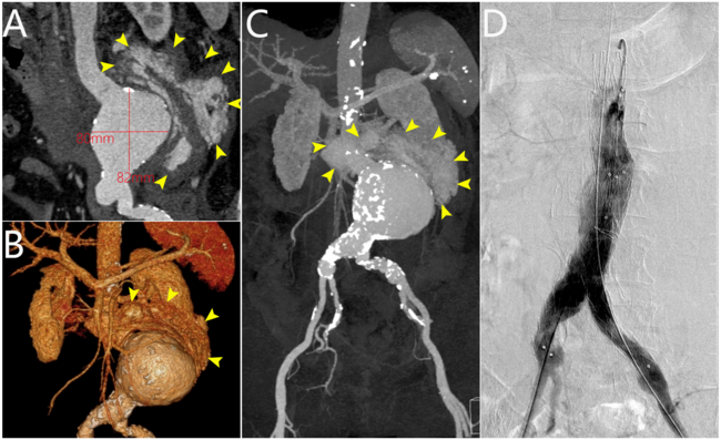
Video 1. Coronary angiography of the saphenous vein (SVG) Y-graft to the first obtuse marginal (OM1) and second diagonal (D2) branch in a right anterior oblique 23°/caudal 27° projection, demonstrating a large aneurysm of the SVG to D2 and a patent SVG to OM1 (proximal to the origin of the aneurysm).
Video 2. Coronary angiography of the saphenous vein Y-graft in a left anterior oblique 20°/cranial 31° projection.
Video 3. Through a left coronary bypass guide catheter, a guide extension catheter was advanced inside the aneurysm sack to allow visualization of the distal vessel segment, demonstrating a small caliber vessel
Video 4. Coronary angiography of the left internal mammary artery graft to the left anterior descending artery in a right anterior oblique 10°/caudal 10° projection, demonstrating possible external compression of the graft by the saphenous vein graft aneurysm.
Video 5. Using a 6F guide extension catheter and a microcatheter, a 0.014-inch workhorse guidewire was advanced through the aneurysm into the second diagonal branch.
Video 6. A 4.0 x10-mm Prestige Plus peripheral vessel coil (Balt) was deployed, with successful occlusion of the outflow segment of the aneurysm.
© 2026 HMP Global. All Rights Reserved.
Any views and opinions expressed are those of the author(s) and/or participants and do not necessarily reflect the views, policy, or position of the Journal of Invasive Cardiology or HMP Global, their employees, and affiliates.