Myopericytoma: What MRI Can Reveal
-
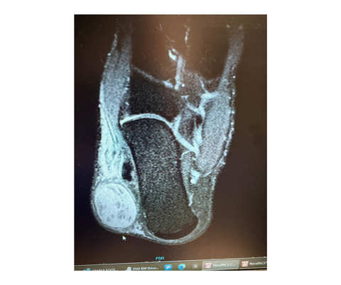
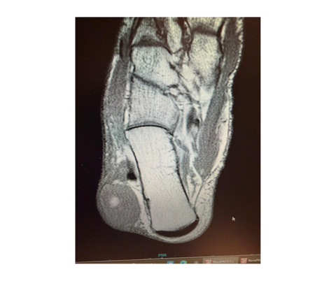
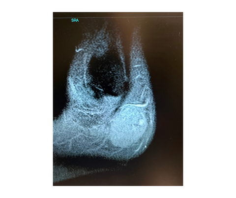
These MRI images represent a case of myopericytoma, a rare, typically benign, slow-growing soft tissue tumor.
-
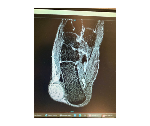
These MRI images represent a case of myopericytoma, a rare, typically benign, slow-growing soft tissue tumor.
-
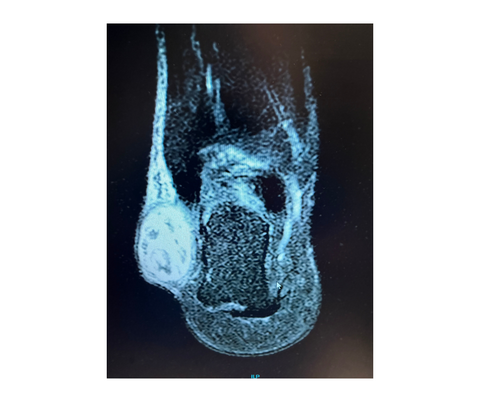
These MRI images represent a case of myopericytoma, a rare, typically benign, slow-growing soft tissue tumor.
-

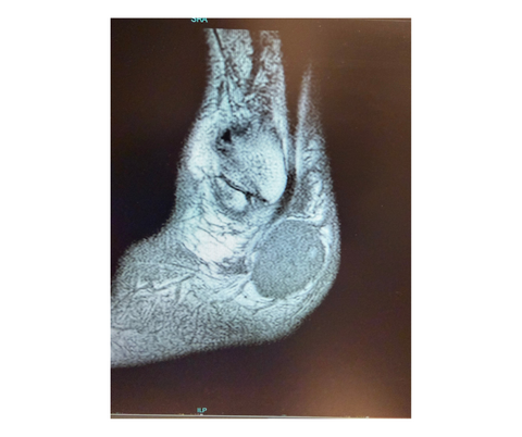
These MRI images represent a case of myopericytoma, a rare, typically benign, slow-growing soft tissue tumor.
-
These MRI images represent a case of myopericytoma, a rare, typically benign, slow-growing soft tissue tumor.
-
These MRI images represent a case of myopericytoma, a rare, typically benign, slow-growing soft tissue tumor.
-
These MRI images represent a case of myopericytoma, a rare, typically benign, slow-growing soft tissue tumor.
Click here for the full feature.
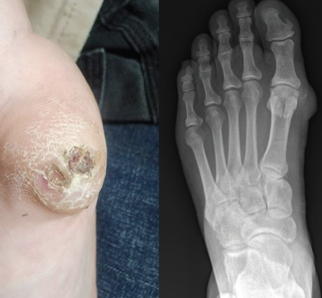
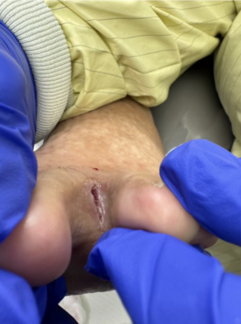
A 40-year-old male of South Asian descent presented to the podiatry office with an enlarging mass in his right ankle, specifically in the subfibular region. The patient reported first noticing the mass approximately 2 years ago. On the initial exam, the patient had palpable pedal pulses and intact neurological status. There were no skin changes noted around the mass. Since then, it had gradually enlarged and began to interfere with his work as a cab driver, specifically with pain when wearing shoes. He described the pain as a mild, dull, throbbing sensation, worsened by movement.
A plain film X-ray of the right foot revealed no significant findings. Subsequent magnetic resonance imaging (MRI) with and without contrast showed a 4.0 x 3.0 x 2.4 cm heterogenous enhancing mass. The MRI interpretation also noted multiple nonenhancing internal components to the mass, a few of which demonstrated an intrinsic hyperintense signal on T1-weighted images.