Phosphaturic Mesenchymal Tumor: Insights on an Unusual Case
When encountering a prominence at the first metatarsophalangeal joint (MTPJ), common considerations include a bunion, bursa, cyst, or even a callus. However, in this case, what initially appeared to be a simple callus at the first MTPJ was ultimately diagnosed as a rare phosphaturic mesenchymal tumor (PMT).
PMTs release fibroblast growth factor 23 (FGF23), which, in excess, can lead to poor bone mineralization and osteomalacia.1 These benign soft tissue tumors typically begin as small, asymptomatic growths, often overlooked due to their slow progression and lack of early irritation or pain.2 However, as they enlarge, they can cause painful, slow- or non-healing ulcers.3 The case described below highlights the importance of considering PMTs in the differential diagnosis of persistent, atypical soft tissue lesions.
Details From the Initial Presentation
A 56-year-old male with a medical history of hypertension and chronic obstructive pulmonary disease (COPD) and a family history of lung cancer (father who worked with asbestos) presented with a mass at the left first MTPJ. He shared that he had initially dismissed the mass as a callus when he noticed it at least 10 years prior. However, it steadily grew in size over the years and began to drain fluid, which motivated him to seek medical evaluation.
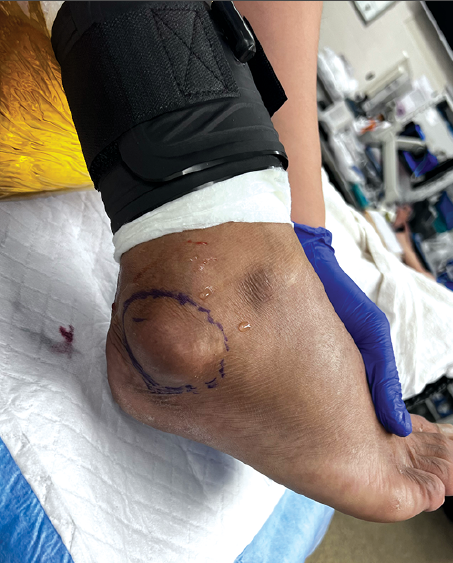
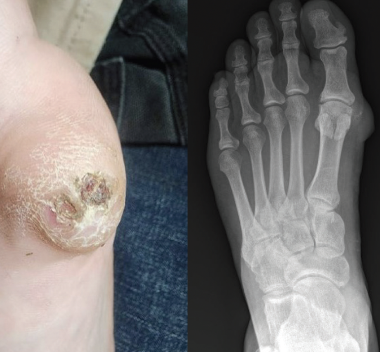
Upon initial examination, the mass appeared as a circular, thickened callus. The area was suspicious for an underlying ulcer due to visible serous drainage and what appeared to be dried heme trapped in the layers of hyperkeratosis. There were no observed irregularities in shape of the lesion or in the overlying skin color. The mass did not appear to interfere with the first MTPJ range of motion and was well defined, firm, and mobile on palpation (Figure 1). Initial plain film radiographs of the left foot showed soft tissue edema at the medial first metatarsal head extending to the MTPJ with some calcification in the soft tissue surrounding the first metatarsal head. There was no evidence of bone erosion or obvious osteophyte growth (Figure 2).
The callus eventually became an ulceration with increased serous drainage from the mass. The ulcer extended over the medial first metatarsal head. The persistent drainage caused maceration, which made healing more difficult. There were no signs or symptoms of infection noted nor did the ulcer probe to bone. However, our team became concerned about the potential for malignancy, as the patient also received a diagnosis of a right posterior shoulder melanoma in the same timeframe and the ulcer over the mass was slow to heal, despite standard local wound care. At this time, it had been a month since the patient initially presented with the mass and a shave biopsy was done for further evaluation and to determine whether the mass was malignant.
Shave biopsy of the ulcerated lesion showed areas of “grungy calcification” with small, monomorphous oval nuclei and stellate cells with thin dendritic processes. FGF23 and phosphate testing helped to further confirm PMT diagnosis. Levels of FGF23 and serum phosphate were 44 pg/mL and 2.5 mg/dL, respectively.
The mass and accompanying ulcer were irritating and painful at times for the patient, however, he related that the pain was intermittent, occurring when the callus reulcerated. As far as course, the ulcer showed a pattern of recurrence, healing to a callus before reulcerating. Approximately one year after the initial exam, the pain had evolved into a constant burning sensation, worsening at night. New radiographs of the foot showed no significant changes from the initial radiographs. Since the mass had become persistently symptomatic, we ordered magnetic resonance imaging (MRI) of the of the foot to help in planning for surgical excision.
Pertinent Perioperative Aspects of the Case
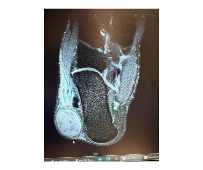
Left foot MRI showed an encapsulated mass measuring 2.6 x 1.4 x 2.4 cm in the plantar subcutaneous fat adjacent to the first MTPJ. The mass abutted the plantar plate and did not extend into the first MTPJ itself. There was no significant joint effusion (Figure 3).
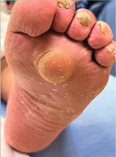
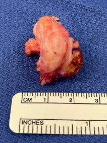
Prior to surgical excision, the mass measured approximately 2.5 cm in diameter (Figure 4). We created a 4-cm incision over the mass to ellipse out the central ulceration. Upon incision and exposure, it was clear the mass was round with well-defined borders. Sharp dissection freed any attachment of the mass to the surrounding skin and careful blunt dissection preserved the mass and its attachment to the now ellipsed central ulcer. The remaining tissue did not have any visible remnants of the mass and appeared to only consist of subcutaneous fat and the intact first MTPJ capsule. The mass itself was firm and springy (Figure 5), which we sent for pathological examination.
The pathology report showed an aggregate of white-yellow adipose tissue with attached white skin measuring 2.1 x 2.1 cm, excised to a depth of 2.3 cm. The attached skin measured 0.6 x 0.5 x 0.2 cm with a brown scaly lesion. Sections of the mass revealed homogenous adipose tissue with necrotic areas. The mass was FGF23 in situ hybridization (ISH) positive, consistent with the diagnosis of a PMT.
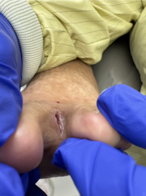
Follow-up postoperatively 3 weeks after mass excision showed a well-healing surgical site with overlying scabbing and no drainage (Figure 6). For a time after suture removal, the patient was lost to follow-up. We were able to have a telephone follow-up with the patient one year later and learned that the mass did not recur and that the surgical site had remained healed.

Discussion of This Unique Type of Tumor
Being very uncommon, PMT can often go undiagnosed. There are a few elements which helped us to arrive at the diagnosis, specifically, laboratory values and pathology.
Clinically, the mass has the characteristics of a benign tumor. It is slow-growing, with a well-defined, rounded shape. As they are oftenn without any specifically malignant symptoms, they can go undiagnosed for years. On average, it can take 5–7 years after the mass is initially noticed for diagnosis to occur.4 The patient in this study received the diagnosis over 10 years after noticing the mass.
Wounds can also form over the mass, which can become chronic. In fact, they may take months to heal, only to recur. In a similar case outlined by Sun and colleagues, the patient had a mass with a chronic, nonhealing ulcer of the medial hallux in which the wound took 8 months to heal. Those authors achieved successful and complete surgical excision after 3 attempts until the mass and its associated wounds did not recur.3 The patient in our report had no recurrence of the tumor or wound after the first surgical excision, although there is evidence of higher recurrence rates of PMTs in elderly patients.5
A key regulator of phosphate homeostasis, excessive FGF23 production leads to phosphate wasting and impaired 1,25-dihydroxyvitamin D synthesis. As PMTs secrete FGF23, this can result in generalized weakened bone formation and osteomalacia.6 FGF23 levels are usually increased in patients with PMT. Although our patient had a level of serum FGF23 within normal limits (reference range ≤59), the FGF23 ISH of the mass itself was positive. Our patient did not show any symptoms of osteomalacia but did show a below normal value of serum phosphate (reference range 2.7–4.5 mg/dL).6
Overall, phosphaturic mesenchymal tumors are often overlooked as they are a rare diagnosis. Although benign, they can significantly impact patients’ quality of life by causing chronic wounds and osteomalacia. Therefore, it is crucial to consider PMTs when evaluating soft tissue masses that do not align with more common diagnoses.
Dr. Kenyon is an attending physician practicing in Modesto, CA.
Dr. Lam is a current third-year resident at Chino Valley Medical Center in Chino, CA.
Dr. Vo is an attending physician practicing in Bakersfield, CA.
1. Jonsson KB, Zahradnik R, Larsson T, et al. Fibroblast growth factor 23 in oncogenic
osteomalacia and X-linked hypophosphatemia. N Engl J Med. 2003;348(17):1656-1663. doi:10.1056/NEJMoa020881.
2. Benson JC, Trejo-Lopez JA, Nassiri AM, et al. Phosphaturic mesenchymal tumor.
AJNR Am J Neuroradiol. 2022;43(6):817-822.
3. Sun X, Ni P, Xie T, Wu S. Phosphaturic mesenchymal tumor along the hallux side inducing a chronic non-healing wound: a case report with literature review. Int J Low Extrem Wounds. 2023;22(4):
779-787. doi:10.1177/15347346221074163.
4. Xiao X, Cai H, Hu S, et al. Phosphaturic mesenchymal tumor mixed connective tissue type and related wound problem: A case report and literature review. Medicine (Baltimore). 2018;97(43):e12507. doi:10.1097/MD.0000000000012507.
5. Sowell J, Srikakolapu S, Preda-Naumescu A, Patel O, Thomley M, Jacobson E, Pavlidakey P. Phosphaturic mesenchymal tumor: A case report and review of surgical outcomes in elderly patients. JAAD Case Rep. 2022;19:34-36. doi:10.1016/j.jdcr.2021.11.002.
6. Bisceglia M, Galliani CA, Orcioni GF, Perrone E, Del Giudice A, Scillitani A. Phosphaturic mesenchymal tumor of soft tissue of the foot: Report of a case with review of the literature. Adv Anat Pathol. 2019;26(5):320-328.