The Convergent Talus Part 3: Mueller-Weiss Syndrome
© 2025 HMP Global. All Rights Reserved.
Any views and opinions expressed are those of the author(s) and/or participants and do not necessarily reflect the views, policy, or position of Podiatry Today or HMP Global, their employees, and affiliates.
Clinical Practice Summary: Mueller-Weiss Disease and the Convergent Talus
Epidemiology & Presentation
-
Rare, often misdiagnosed condition affecting the navicular bone
-
Typically occurs in middle-aged women (~40s)
-
Commonly mistaken for tibialis posterior dysfunction (TPD)
Pathophysiology
-
Involves avascular necrosis of the navicular
-
Blood supply rich at the tuberosity, but central/lateral navicular vascularity is tenuous
-
Trauma or overuse may disrupt radial vessels, leading to bone fragmentation and collapse
Radiographic Features
-
AP view: “Comma-shaped” navicular due to collapse
-
Lateral view: Dorsal bone fragmentation, misread as accessory ossicle
-
Progressive talar shift: Forward, downward, and lateral displacement causing convergent talus
Deformity
-
Creates paradoxical pes planovarus:
-
Heel varus from lateral talar shift
-
Flatfoot appearance from talar descent into navicular defect
-
Clinical Pitfalls
-
Misdiagnosed as TPD flatfoot → inappropriate fusion procedures → risk of nonunion
-
Misinterpreted dorsal bone as excess ossification → unnecessary resections
Staging & Surgical Options (Maceira Classification)
-
Stage 1: Early varus heel and talar convergence
-
Valgalizing calcaneal osteotomy may correct alignment, relieve stress, and allow consolidation
-
-
Stage 2: Comma-shaped navicular, talonavicular involvement only
-
Partial naviculectomy + talonavicular fusion possible
-
-
Stage 3: Severe collapse, transcuneiform joint involvement, varus heel overloads calcaneocuboid joint
-
Options: Cuneiform fusion, triple arthrodesis (subtalar, calcaneocuboid, cuneiforms)
-
-
Stage 4: Advanced deformity with ankle involvement, talar-tibial impingement
-
May require pantalar fusion, or complex reconstruction with Charcot triple + supramalleolar osteotomy + fibular transposition
-
Key Takeaways for Practice
-
Early recognition is critical—intervene in Stage 1–2 before midfoot and rearfoot arthritis develop
-
Always evaluate for varus heel with flatfoot—hallmark of paradoxical pes planovarus
-
Misdiagnosis risks failed procedures and worsened disability
-
Treatment becomes increasingly complex and morbid with progression
Transcript
Jennifer Spector, DPM: Welcome back to Podiatry Today podcast where we continue to bring you the best and foot and ankle medicine and surgery from leaders in the field. I'm Dr. Jennifer Spector, the Assistant Editorial Director for Podiatry Today, and we're continuing our series with Dr. Visser on the convergent talus. This time we're going to get into an uncommon condition, Mueller-Weiss syndrome, and how this impacts the concept of the convergent talus.
Dr. Visser was the president of the St. Louis Podiatric Medical Society and is a past president of the Missouri Podiatric Medical Association. He was an examiner of the American Board of Podiatric Surgery and served on the Missouri State Board of Podiatric Medicine over several governorships. He's been a residency director for four decades, most currently at SSM DePaul, Foot and Ankle Reconstructive and Trauma Surgical Program in St. Louis. As part of this program, he has currently trained 114 residents. In 2015, he was inducted into the Kent State University School of Podiatric Medicine Hall of Fame.
There's another condition that you've mentioned before, that I think a lot of DPMs may not know as much about. And it relates to the convergent talus as well. And that's Mueller-Weiss disease. Could you get into that a little bit for us?
H. John Visser, DPM, FACFAS: Yeah, so Mueller-Weiss disease is a condition that is not what I would call highly common, but it is another one of these conditions that a lot of people misdiagnose it. It is very commonly misdiagnosed in the human foot. And basically what happens is, is that the patient is usually female, usually in their mid-40s. And it kind of falls into that group of tibialis posterior dysfunction, and oftentimes it will get misdiagnosed. And I've had interesting case presentations with the residents, with attending staff, where literally this condition was treated as a form of TP dysfunction, but they failed to recognize that the condition was Mueller-Weiss disease.
Now, what this condition is—it affects the navicular bone. And how it affects the navicular bone is we have to kind of review the blood supply. We know the blood supply to the tubercle of the navicular is very rich. We have a big cascade of arterial vessels in and around the tuberosity of the navicular bone with coming from the medial plantar artery and the posterior tibial artery. The blood supply from there becomes relatively tenuous. We have three radial vessels that then pass from medial to lateral across the body of the navicular, the so-called central and lateral poles. So if there's anything, usually, it is traumatic in nature, and it can be from overuse. It can be from an incidence of a fall where the patient really didn't sustain a whole lot of injury at the time, where a fracture can occur, what we call an [inaudible] fracture, that basically goes perpendicular to the body and cuts off the three radial blood supplies. And again, the true etiology has never been really finally determined, but it is believed to be some form of a traumatic event. Once this occurs, avascular necrosis of the navicular begins to occur. And what happens is, is that the central and lateral body will begin to fragment and break down.
Jennifer Spector, DPM: So what might somebody see on x-ray for this?
H. John Visser, DPM, FACFAS: On the AP view, what you will begin to see is what we call the development of a comma-shaped navicular bone. And what happens as this progresses, this basically avascular necrotic process, the talus will begin to fill the void loss because what's happening is the AP diameter of the navicular bone becomes smaller and smaller and narrower and narrower as the comma forms. So as it does this, the talus begins to shift forward and you begin to get a defect in the plantar portion of this interface. And as the disease, and there's been a classification described radiographically, the Maceira Classification, as it gets into the stage 3 and 4, literally you just have this comma thin of a vascular necrotic bone in the central medial column, the talus has moved forward and it has moved downward. It also has shifted lateralward. It shifts lateralward and this is the convergent issue and it creates what's called closed kinetic chain supination to the rearfoot. And this will then produce what has been referred to as a paradoxical pes planovarus foot.
Jennifer Spector, DPM: Paradoxical—tell us more about that.
H. John Visser, DPM, FACFAS: Now, that's kind of a long term there, but basically what happens is, is that the heel varus is created by the fact that the talus has shifted lateralward. And as this size of the central lateral pole in the navicular becomes decreased in size, it's just lateralward and it supinates the subtalar joint. That's the varus part. That's where you come up with the varus. The flatfoot is created by the fact that the talus had moved forward and falls down into this defect created by loss of volume of the central body of the navicular bone. And so the arch will look like it's collapsed. That you're walking on the mid arch of your foot, yet you have a varus heel. And that's where the term paradoxical pes planovarus deformity basically occurs in this particular situation. Now, again, depending on the stages of it, determine oftentimes what you will do from a surgical standpoint. In the very early stages, okay, stage 1, again, what happens is you're beginning to see the varus heel.
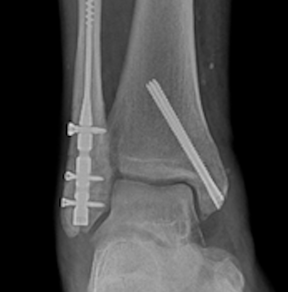
The interesting thing too is on the lateral view you will see all these fragmentations of bone dorsally over the midfoot and this is all created by this abascular bone being broken up and this is what happens. The patient comes in. The surgeon notices on the lateral view, this fragmentation, they then think they've got an excess ptosis on the top of the navicular bone. This is a very painful condition. And so they assume, well, let's just go in and we'll go ahead and resect this redundant bone. And I had a patient that had this done two times before I saw her and made the diagnosis that she had a Mueller-Weiss disease condition, an avascular necrosis of the navicular bone. They fail to recognize that the navicular had undergone an avascular necrotic process. Same thing happened with this TP dysfunction foot. It was a patient, similarly, fragmentation, yet the patient had what they perceived a flat foot, but they didn't really look closely to see that they had a varus heel. They didn't really look also at the divergence/convergence issue of the talus. Clearly, the talus was convergent. They just didn't look and just assumed it was divergent, and then went ahead and did a TP dysfunction repair, which actually included a tail navicular fusion, which you know went on to a nonunion because they did not recognize that the fact that the navicular had avascular necrotic changes.
But in a stage 1 situation where in the very first phase, as you get the lateral translation, the convergence of the talus on the navicular is, it's beginning to lose some of its bone content. The heel tips into varus. If you can catch it at that early stage, you can do a valgalizing calcaneal osteotomy. And when you perform that, you are basically going to shift the talus from its convergent position to a divergent condition. And in that way, with a simple calcaneal osteotomy, you'll avert the stress on the central lateral pull of the navicular and basically allow the avascular necrotic issue to go through its consolidation process.
Jennifer Spector, DPM: As it progresses, what can we expect to see then?
H. John Visser, DPM, FACFAS: As it gets into the stage 2, we begin to start to see more of the comma shape. We have the varus heel occurring, but at this point, the cuneiform bones, which it articulates with distally, are still not highly involved, but we're dealing highly in this particular situation with talonavicular disease. And we have the options of doing a near navicular here, we can actually remove the diseased portion of the central lateral pole of the navicular and do a fusion of the talonavicular joint because we still have a large portion of the navicular present that's supplied by the medial plantar artery and the posterior tibial artery. That defect can be just filled with nothing. You can put an allograft in there with you like, but the fusion process would be centered at the talonavicular joint. When you get into the stage 3, there you're getting a real true comma sign that most people should be able to diagnose.
The problem is the first two stages where there's failure oftentimes recognition, in this case, that what do we've got? We've got a situation now, not only is the talonavicular involved, we also have the transcuneiform joints, okay? And basically, again, we have that various heel situation that's been created. And we have this foot that basically appears flattened. So we're gonna have to attempt to effuse the cuneiforms to remaining navicular bone. He usually use some type of an autoallograft to get that lateral portion. The other problem that sometimes occurs is is the calcaneal cuboid joint is overloaded by this varus position of the subtalar joint. Just a reminder, whatever the subtalar joint does, the calcaneal cuboid joint does. So if the subtalar joint is in a varus supinated position, so is the calcaneal cuboid joint. So in some instances, you're going to have to consider a full triple arthrodesis being done where you basically fuse the subtalar joint to get it out of that varus position, the calcaneal cuboid joint, which is overloaded in a varus state. And then you have to fuse the so-called, what we call a Charcot type of triple, which includes the cuneiform bones. You can also consider what's called a [inaudible], which is where you excise the complete body of the navicular what's left and fuse it to the cuneiform bones, which requires shortening of the calcaneal cuboid side. But this is not usually well recommended.
Jennifer Spector, DPM: As this continues to get worse in severity, what comes next?
H. John Visser, DPM, FACFAS: Then you get into your stage 4 in the most severe form and this can begin to affect the ankle. Remember what happens is as that subtalar joint gets into a more severe varus state, it will have an effect on the position of the talus and you will end up with impingement basically with the talus in the medial area between the tibia and the talus medially. It'll be in a varus, intrinsically varus position along with the heel. In that particular instance, you may have to consider a a pantalar fusion, or certainly you may be able to combine a so-called Charcot triple with a supramalleolar osteotomy and opening wedge of the tibia along with a transpositional osteotomy of the fibula. So you can see how complex this can get, and the thing is a very, a painful problem. And so it often requires treatment. The key is to get it within the first two stages. You want to try to avoid where you have transcuneiform and talonavicular arthritis, because what happens with this is you begin to have changes in the rearfoot that are significant that also affect the midfoot.
Jennifer Spector, DPM: Thank you so much, Dr. Visser, for sharing this with us today, and I know there's a lot more to say on the convergent talus, so be sure to stay tuned for future episodes of Podiatry Today podcast on your favorite podcast platforms and on podiatrytoday.com.