Bridging Disciplines in Lower-Extremity Trauma
A Podiatrist’s Intraoperative Perspective on Ankle Fracture Fixation
A Podiatrist’s Intraoperative Perspective on Ankle Fracture Fixation
© 2025 HMP Global. All Rights Reserved.
Any views and opinions expressed are those of the author(s) and/or participants and do not necessarily reflect the views, policy, or position of Podiatry Today or HMP Global, their employees, and affiliates.
In an era of increasingly complex lower-extremity injuries, interdisciplinary collaboration between orthopedic surgery and podiatry is critical for optimizing patient outcomes. While podiatric surgeons are extensively trained in foot and ankle pathology, opportunities to participate in higher-level trauma cases—particularly ankle fractures extending into the tibia—remain limited in some academic settings. However, in such settings where podiatric involvement in trauma is not currently robust, this case presented below provides an example of an intentional effort to bridge that gap and begin to foster relationships that drive the profession forward.
This report presents the unique perspective of a podiatric physician integrated into the surgical management of an unstable ankle fracture, performed by an orthopedic trauma surgeon. The experience highlights both the technical nuances of fracture care and the collaborative potential of a multidisciplinary limb preservation approach.
Case Description
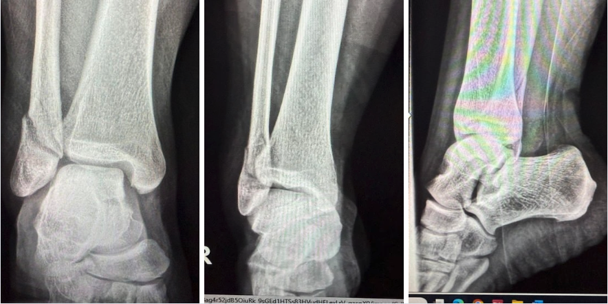
The patient was a healthy 39-year-old female who sustained a closed distal fibular fracture (Weber B type). After confirming the diagnosis radiographically and completing preoperative assessments, the team obtained informed consent. Risks reviewed included infection, bleeding, nerve injury, hardware failure, stiffness, the need for further procedures, and anesthesia-related complications, including rare, but serious, outcomes such as thromboembolism, myocardial infarction, and death. The surgeon appropriately marked the surgical site in the preoperative holding area.
Using tourniquet control and a lateral incisional approach to the fibula, careful dissection through subcutaneous tissues exposed the periosteum. A longitudinal incision of the periosteum revealed the fracture site, which the surgeon debrided and irrigated. A Weber clamp assisted with fracture reduction, confirmed intraoperatively using AP and lateral fluoroscopic views.
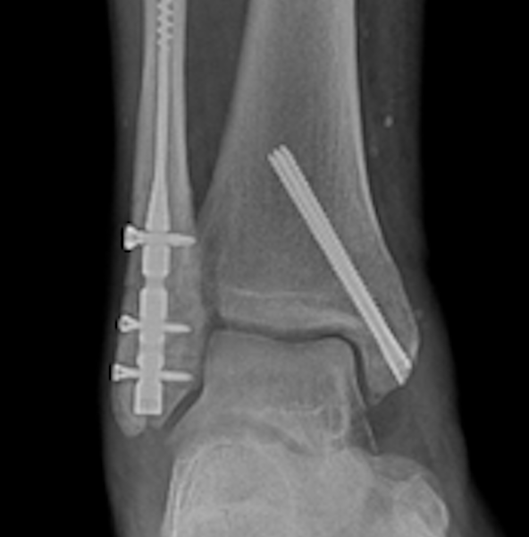
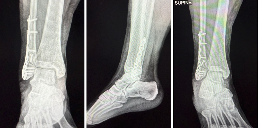
Fixation included a single lag screw to stabilize the long oblique fracture. A 3-hole distal anatomical fibular plate provided neutralization. Two bicortical screws inserted into the fibular shaft secured the plate proximally, and 3 locking screws affixed the distal fragment. Fluoroscopy allowed for verification of all screw positions.
Following thorough irrigation, the surgeon confirmed hemostasis after tourniquet deflation. Layered closure was with 2-0 Vicryl for the deep tissues, followed by 2-0 Vicryl for the subcutaneous layer and nylon for the skin. Application of a sterile dressing and immobilization with a CAM boot concluded the case.
The postoperative plan for this patient included:
- Touch-down weight-bearing on the right ankle for 6 weeks
- Limb elevation encouraged
- Dressing change scheduled in one week with a physician assistant
- Follow-up appointment in 2 weeks
- Anticoagulation: enoxaparin sodium (Lovenox) subcutaneously for 2 weeks, followed by aspirin 81 mg twice daily for 4 weeks
Intraoperative Observations
As a podiatric physician participating in the procedure, I observed the technique and decision-making involved in distal fibular fracture fixation from the orthopedic trauma perspective. Notably, I felt that the use of a neutralization plate combined with both bicortical and locking screws reflected a thoughtful approach to optimizing stability in an active, weight-bearing adult.
I observed that the fracture exposure was efficient and well-controlled, allowing clear visualization for reduction. Bicortical lag screws provided compression across the fracture site, while locking screws reinforced fixation in distal segments with less bone stock.
The combination of construct mechanics appeared offer a reliable method to protect against rotational and shear forces during the early postoperative phase. This surgical experience provided valuable insights into fixation principles applicable to a range of foot and ankle procedures, particularly in managing trauma in otherwise healthy adults.
Interdisciplinary Value
This case underscores the clinical and educational value of interdisciplinary collaboration between podiatry and orthopedic surgery. Scrubbing into this procedure offered a deeper understanding of fracture fixation strategies and reinforced the importance of unified care pathways. It also highlighted opportunities for improved communication and shared decision-making when managing traumatic injuries involving the foot and ankle.
From a systems perspective, the integration of podiatric physicians into orthopedic trauma cases can enhance continuity of care, especially in the postoperative period where wound surveillance, offloading strategies, and biomechanical assessments are essential. It promotes cross-specialty learning and strengthens institutional efforts to build cohesive, outcome-driven teams.
Such experiences can inform future collaboration in developing limb preservation protocols, co-management models, and resident training initiatives that embrace a truly multidisciplinary approach to musculoskeletal health.
Future Directions
The collaboration between podiatry and orthopedic surgery in this case offers a foundation for future interdisciplinary initiatives, particularly in rural, underserved communities like Dansville, New York, where access to specialized musculoskeletal care is limited. One key area for development is the creation of formalized co-management protocols for lower-extremity trauma, enabling more seamless coordination between surgical teams, wound care providers, and rehabilitation services.
Additionally, the institution could benefit from joint educational programs that expose residents, medical students, and allied health professionals to the principles of collaborative fracture care and limb preservation. These programs would not only enhance clinical competency but could also strengthen the multidisciplinary culture needed to manage complex pathology in resource-constrained settings. In the long term, these collaborative efforts may support the development of a regional center of excellence in limb preservation, anchored in shared surgical education, research initiatives, and patient-centered care models tailored to the needs of rural populations.
Conclusion
This case serves as a compelling example of how shared operative experiences between podiatric and orthopedic surgeons can drive clinical insight and system-level innovation. Beyond the technical knowledge gained, the opportunity to collaborate in real time fosters mutual respect and highlights the complementary strengths of each discipline.
In underserved areas such as Dansville, New York, these collaborations are not only educational—they are essential. By breaking down specialty silos and promoting joint responsibility for patient outcomes, healthcare teams can deliver more comprehensive, timely, and effective care. Ultimately, this experience reinforces the importance of interdisciplinary engagement as a cornerstone of both clinical excellence and health equity, especially in settings where resource limitations make cohesive teamwork not just beneficial, but imperative.
This case highlights a developing collaborative model in which podiatrists, particularly in rural or resource-constrained environments, seek opportunities to co-manage trauma cases traditionally reserved for orthopedics. While formal training in ankle fracture ORIF is not universally limited for DPMs, institutional protocols, surgical privilege structures, and call responsibilities often dictate the initial assignment of these cases. In this scenario, my involvement reflects an intentional effort to bridge disciplinary silos, enhance perioperative care continuity, and contribute to an integrated limb preservation model. This interdisciplinary approach may serve as a template for similar underserved or mid-sized institutions where care teams must adapt to limited subspecialist availability.
Dr. Archer is a board-certified podiatrist and certified wound specialist. She serves as a Senior Instructor in the Department of Orthopaedics at the University of Rochester Medical Center. Her clinical expertise includes diabetic foot care, limb preservation, and wound management, with a strong focus on healthcare delivery in underserved and rural communities.
Dr. Al-Hardan is an Assistant Professor of Orthopedic Surgery at the University of Rochester Medical Center. His clinical practice focuses on orthopedic trauma surgery, with interests in interdisciplinary surgical care, joint preservation, and improving musculoskeletal outcomes in rural populations.