Case Report

Case Study of Limb Salvage in a Complex Wound With Necrotizing Fasciitis and Osteomyelitis

© 2025 HMP Global. All Rights Reserved.
Any views and opinions expressed are those of the author(s) and/or participants and do not necessarily reflect the views, policy, or position of Podiatry Today or HMP Global, their employees, and affiliates. 

Necrotizing fasciitis with concurrent osteomyelitis represents one of the most formidable challenges in limb preservation, particularly in patients with multiple comorbidities. This case study highlights how aggressive surgical debridement, multidisciplinary coordination, and staged reconstruction enabled successful limb salvage in a high-risk diabetic patient who declined amputation.

Key Clinical Takeaways

1. Early, aggressive, and repeated debridement is essential for survival and limb preservation.

Prompt recognition of necrotizing fasciitis and osteomyelitis—supported by tools such as the LRINEC score—and rapid surgical intervention remain critical. Multiple staged debridements were required to control infection and prevent further tissue destruction in this high-risk case.

2. Limb salvage can be achievable even in patients with significant comorbidities when multidisciplinary care is applied.

Despite the patient’s diabetes, ESRD, HIV, peripheral vascular disease, and severe infection burden, coordinated care among podiatric surgery, infectious disease, and vascular teams enabled infection control and functional limb preservation.

3. Staged reconstruction with NPWT, skin substitutes, and split-thickness skin grafting supports durable closure after infection control.

Following clearance of infection, the combination of negative pressure wound therapy, skin substitutes, and STSG facilitated granulation and ultimate wound closure, allowing the patient to progress to full ambulation without recurrence.

Necrotizing fasciitis and osteomyelitis are emergent and life-threatening presentations commonly encountered when taking foot and ankle call, particularly in patients with diabetes. These conditions often require urgent surgical intervention—typically emergent incision and drainage versus amputation—alongside aggressive antibiotic therapy.Necrotizing fasciitis is a rapidly progressing soft tissue infection that spreads along fascial planes, often exploiting areas of poor vascular supply.1 While it remains primarily a clinical diagnosis, the Laboratory Risk Indicator for Necrotizing Fasciitis (LRINEC) score is frequent tool clinicians use to support diagnosis.2 Mortality rates for this condition remain alarmingly high, ranging from 20% to 80%.1

Osteomyelitis in the diabetic foot has a similar poor prognostic association. The presence of osteomyelitis increases the risk of amputation by a factor of 4 in diabetic foot wounds.5

Furthermore, outcomes following major amputations are grim: the 5-year survival rate is approximately 47.9% for above-knee amputations and 31.5% for below-knee amputations.6

Given the high morbidity and mortality associated with both necrotizing fasciitis and diabetic foot osteomyelitis, it is imperative that foot and ankle surgeons are well-versed in the diagnosis and management of these conditions.

In this piece, we present a case of successful limb salvage in a high-risk patient with diabetes and concurrent necrotizing fasciitis and osteomyelitis—a scenario that typically portends a poor prognosis.

Key Case Details

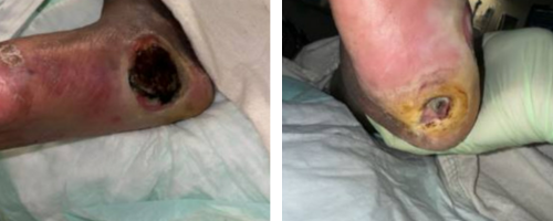
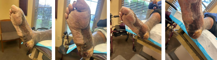
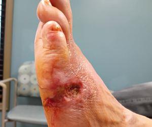
A 52-year-old African-American female presented to the emergency department with worsening wounds on her left foot. Her past medical history included type 2 diabetes, human immunodeficiency virus (HIV), end-stage renal disease on dialysis, anemia, hypertension, a history of MRSA infection, peripheral neuropathy, and peripheral vascular disease. She had been undergoing prolonged outpatient wound care at another institution but developed worsening malodor and swelling over the past 24 hours (Figures 1 and 2). 

Initial lab results revealed white blood cell count (WBC) >22,000 cells per microliter, hemoglobin <10 g/dL, blood glucose >280 mg/dL, creatinine 3.1 mg/dL, CRP >250 mg/L, and sodium 126 mmol/L, consistent with a LRINEC score of 12—highly suspicious for necrotizing fasciitis. X-ray and computed tomography (CT) imaging confirmed extensive soft tissue gas throughout the plantar and medial aspects of the foot. The team placed a consultation for infectious disease, and the patient began broad-spectrum intravenous antibiotics.

A thorough discussion with the patient and her family reviewed the options of below-knee amputation versus attempted limb salvage via incision and drainage with bone biopsy and negative pressure wound therapy (NPWT) application. Despite understanding the guarded prognosis, the patient declined amputation.

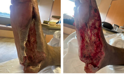
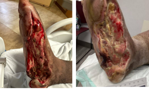
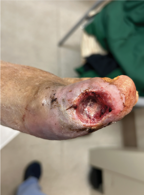
We emergently took the patient to the operating room for the stated attempt at limb preservation. On postoperative day 1, we noted ischemic skin flaps during the dressing change, and planned for repeat debridement (Figures 3 and 4).

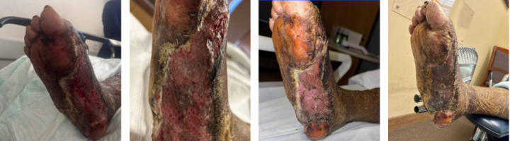
Although the WBC trended downward, it rose again on postoperative day 4, prompting an urgent repeat CT scan, which again showed plantar soft tissue emphysema (Figure 5). This resulted in a third surgical debridement.

Following this, the patient's WBC normalized. During her admission, arterial studies demonstrated significant peripheral vascular disease. Vascular surgery recommended below-knee amputation given the combination of infection and ischemia. However, we continued limb salvage efforts per the patient’s wishes. Pathology confirmed osteomyelitis of the foot and calcaneus on all procedures. The patient was discharged on intravenous antibiotics per the infectious disease team and NPWT with outpatient follow-up plans.

Here one can see the preoperative appearance of the left foot.
Figures 1 and 2. Here one can see the preoperative appearance of the left foot.


These photos show the preoperative necrosis (left) and the foot's appearance after removal of necrotic tissue (right).
Figures 3 and 4. These photos show the preoperative necrosis (left) and the foot's appearance after removal of necrotic tissue (right).


Outpatient Management

The patient was followed closely in the outpatient clinic. Weekly serial debridements were performed along with wound VAC therapy. Approximately one month after her initial debridement, operative debridement and application of a skin substitute graft were performed (Figure 6). One month later, a staged split-thickness skin graft (STSG) was harvested and placed. Preoperative cultures revealed only rare bacterial growth, and appropriate antibiotics were administered pre- and post-operatively based on pre-operative and operative culture results. Over 90% of the STSG successfully incorporated shortly after surgery (Figures 7, 8, 9, 10).

Local wound care and weekly debridements continued (Figure 11,12). The patient progressed well and was transitioned to weight-bearing in a CAM boot. By six and a half months post-STSG, complete healing was achieved with no recurrence or breakdown noted (Figure 13,14). She was ambulating independently at this time.

Interim photos between surgical procedures left foot.
Figures 5 and 6. In the left image one can see the clinical appearance prior to the third incision and drainage. The image on the right depicts the appearance prior to the skin substitute graft. 


Early incorporation of the split thickness skin graft.
Figures 7-10. From left to right, these photos show incorporation of the split thickness skin graft at 2 weeks, 2 weeks, one month, and 9 weeks.


Continued progress throughout several procedures.
Figures 11-14. These photos from left to right show progress from 6 to 6.5 months postop split thickness skin grafting.


Final Management Notes

This case highlights the challenges and potential for successful limb salvage in patients with necrotizing fasciitis and osteomyelitis. It underscores the importance of thorough surgical debridement, staged reconstruction with skin grafting, and consistent local wound care. Multidisciplinary management in these high-risk cases may prevent amputation and substantially improve quality of life and long-term outcomes in patients with diabetes.

Dr. King is an Associate of the American College of Foot and Ankle Surgeons.

Dr. De Guzman and Dr. Cohen are Residents at East Liverpool City Hospital in East Liverpool, OH.

Dr. DiDomenico is a Fellow of the American College of Foot and Ankle Surgeons and Fellowship Director at NOMS Foot and Ankle Center.

References

1.    Wallace HA, Perera TB. Necrotizing Fasciitis. [Updated 2023 Feb 21]. In: StatPearls [Internet]. Treasure Island (FL): StatPearls Publishing; 2025 Jan-. Available from: https://www.ncbi.nlm.nih.gov/books/NBK430756/
2.    Wong CH, Khin LW, Heng KS, Tan KC, Low CO. The LRINEC (Laboratory Risk Indicator for Necrotizing Fasciitis) score: a tool for distinguishing necrotizing fasciitis from other soft tissue infections. Crit Care Med. 2004;32(7):1535-41. doi: 10.1097/01.ccm.0000129486.35458.7d. PMID: 15241098.
3.    Wong CH, Chang HC, Pasupathy S, Khin LW, Tan JL, Low CO. Necrotizing fasciitis: clinical presentation, microbiology, and determinants of mortality. J Bone Jt Surg Am. 2003;85-A:1454–60.
4.    Fontes RA Jr, Ogilvie CM, Miclau T. Necrotizing soft-tissue infections. J Am Acad Orthop Surg. 2000;8:151–8.
5.    Mutluoglu M, Sivrioglu AK, Eroglu M, et al. The implications of the presence of osteomyelitis on outcomes of infected diabetic foot wounds. Scand J Infect Dis. 2013;45(7):497-503. 
6.    Subramaniam B, Pomposelli F, Talmor D, Park KW. Perioperative and long-term morbidity and mortality after above-knee and below-knee amputations in diabetics and nondiabetics. Anesth Analg. 2005;100(5):1241-1247.