Slippers Gangrene: A Deeper Dive Into a Unique Case
© 2025 HMP Global. All Rights Reserved.
Any views and opinions expressed are those of the author(s) and/or participants and do not necessarily reflect the views, policy, or position of Podiatry Today or HMP Global, their employees, and affiliates.
Click here to read the case study.
Transcript
My name is Babajide Ogunlana. I'm a podiatrist practicing out of Missouri City, Texas.
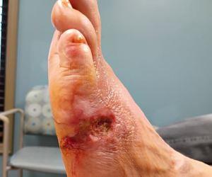
So this was actually a unique presentation just by virtue of the way it looked. So I've had cases where we've had digital gangrene or forefoot gangrene, but this particular case was a bilateral case, but one of them actually looked like a slipper. Basically, like you put on your foot into a slipper. And that's exactly how it presented on the left foot while the right foot was the typical forefoot gangrene involving all the toes. So it was pretty unique in that sense, just the way it looked.
And I tried to search in the literature to see how many cases are out there that have been reported and I didn't see that many, so I thought it was unique. And the fact that it was also wet in this case, it came into the office foul smelling. And it had been going on for several months, but he apparently went home, was discharged and was doing his regular showers and the dry gangrene was getting wet and then it converted to wet gangrene, which over time was just not a good then and it involved the entire plantar aspect of the foot all the way to the forefoot. So having to surgically operate on it was quite a challenge, because to get tissue coverage was a big challenge.
Every time I've seen these types of cases where you have forefoot gangrene, sudden onset, it's usually been people in the ICU setting that have been on pressure therapy. So in that case, what that means is they've had cases of hypertension, severe hypertension, and they've had to be in the ICU. They've had to be put on pressure therapy. And so as a result of that, usually what that means is the body's trying to basically maintain the blood pressure so it preserves the blood supply to the rest of the trunk or the body and then stabs the extremities. So that's basically what ends up happening is sometimes it's in the fingers and oftentimes it's in the toes or the foot and in this case it was bilateral.
Addressing the Challenge of Limb Preservation and Tissue Coverage
I immediately admitted him to the hospital. The plan was to do transmetatarsal amputation open because this was wet gangrene on both sides. We had x-rays done and there was gas bubbles. The entire plantar aspect of the left foot, including the forefoot and on the right side on the forefoot, where the gangrene was.
What you noticed was, you had fully demarcated gangrene of the forefoot to the midfoot area. However, on the boundary between the normal skin and the gangrenous area, you actually saw red tissue, grand lesion tissue. So this patient did not have the typical case of peripheral vascular disease where there's a major vessel occlusion. It was mostly like a thromboembolic episode that really blocked the vessels, the blood supply to the area of the forefoot. So even when you do arterial Dopplers, most of it is mild to moderate, it's not like severe stenosis or occlusion. And in this case, the vascular team did not have to do anything. Infectious disease internal medicine did a multidisciplinary management for this patient.
So surgical decision was made because it was an emergent case. So I admitted him to the hospital, took him to the OR that same night. Bilateral open TMAs. The right side again was forefoot gangrene, so was able to do it up until the midfoot and just left it sort of like a guillotine-type amputation. While the left side was more involved.
Now on both of them I used tourniquet. Again, what I was saying about not your typical stenosis or peripheral vascular disease type of case. I used tourniquet, so I was able to basically peel off the left foot, forefoot, midfoot, and then the plantar aspect—it literally came apart. The slippers literally was on the table, and then you could expose the entire structure on the bottom of the foot on the left side. So all the intrinsic extrinsic muscle, you know, belly, bones, I did a parabola resection for the transmetatarsal and then I let down the tourniquet and I can tell you the blood supply was just gushing so that tells you the blood supply is adequate it was just you needed to get rid of all the infected dead tissue and that's what we did.
So now I turned back on the tourniquet, and then I put some moisture control dressings because it was severely infected. So, it wasn't time for closure yet, it was more of just prepare and then do dressing changes over the next few days, and then start thinking about closure or how we're going to close this. And that was the challenge. What do we use? And we had to, about three to four days later, took it back, and at this time. So we were using some moisture control dressings on a daily basis and it was highly exudative, a lot of drainage. But the wound bed was a lot cleaner. So day one, day two, day three, the smell—gone.
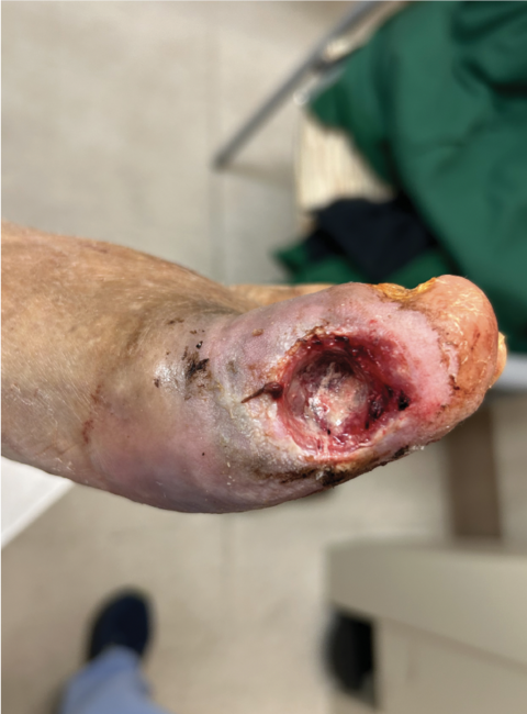
But now we have large wounds on bilateral feet. So we decided to use some advanced healing wound care type products and over the next four weeks, five weeks, that right side closed nicely, was able to fill in with granulation tissue and got some epithelialization and the skin basically grew in to cover the deficit. On the left side, used the same thing, but again, it's a larger area, so we had to use a just sheet of the cytolde and then the micrometrics are powders on it. Again, staple it. Usually when we staple it's able to secure the graft in place and I made a decision not to use a negative pressure on it. I mean it was well adherent and I had it stapled and then I had the bulky dressing. The amount of drainage was excessive so I just used a moisture control dressing on a daily basis while he was in the hospital, and then once he got discharged, we saw him in the office once a week.
He was strictly non-weight-bearing to the left side, as you can imagine, and on the right side, he could bear weight on the heel, as tolerated. He used a rolling knee walker to move around, and he did well. It took a while. The right side healed quickly. The left side, again, as you can imagine, we literally had to grow tissue, skin, to cover the entire plantar foot that was remaining.
And the question could be asked, why did we not maybe put a native skin graft or autologous graft-like tissue. This is on the plantar aspect of the foot. I've had cases of patients that have had skin grafts taken from their thigh and placed on the bottom of the foot—they look different. And there's always an issue with the interface. Every time they come in they develop some hyperkeratosis along the edges that has to be shaved out periodically; otherwise it's just painful. And I just wanted to see if we can recruit the native skin or tissue to grow over the deficit. And I can tell you, we did and it's pretty impressive.
Pertinent Pearls From the Case
You know in some settings there will be automatic BKA. I think it's more of a comfort level issue and this patient was not even diabetic, remember. Again his other comorbidities were aortic aneurysm. He had surgery for that and then he had complications and anytime somebody comes to me with a sudden onset of gangrenous changes, not just one toe but multiple toes, I usually ask them, "Were you in ICU? Were you, you know, intubated, you know, on pressure therapy? Did you have severe hypertension?" And majority of the time, yes.
There are things that we could do to save the limb. I mean, we shouldn't just always rush to leg amputation because if you take off one leg, usually within a few years the other leg is gone and then they die within five years. So, it's worth it to try to preserve the limb.