


Distal Dorsal Radial Access: An Experience Worth Sharing
© 2025 HMP Global. All Rights Reserved.
Any views and opinions expressed are those of the author(s) and/or participants and do not necessarily reflect the views, policy, or position of Cath Lab Digest or HMP Global, their employees, and affiliates.
Ananth N. Kumar, MD, FACC
Clark Regional Medical Center, Winchester, Kentucky
Dr. Ananth Kumar can be contacted at heartmatterpsc@gmail.com.
Click here for a PDF of this article. Logging in or registration may be required (it's free!).
Radial access has changed the landscape of complications during cardiac catheterization. The push for radial access has been accepted by a majority of cardiologists, with approximately 60% of coronary angiograms across the country now being performed through this access.1 The touted advantages of the radial access are patient comfort, reduced access site complications, and reduced mortality and morbidity post angiography. It has greatly improved post-procedure limitations for patients and post-procedure complication anxiety for interventionalists.
However, radial access has had its own unique issues. Arterial spasm with difficulty in maneuvering catheters, leading to abandonment of the access site, is a real occurrence. Also, there is a historical incidence of radial artery closure of 7.7%.2 While radial access success has been reported in the 90%+ range, there is still an occurrence of crossover to femoral access in 3-10% of patients for various reasons, including but not limited to difficulty in engagement, support, subclavian occlusions, and tortuosity issues.3
From a patient perspective, the discomfort of external rotation with dorsiflexion of the wrist during the procedure and prolonged high pressure post-procedure for hemostasis are well known. Post-procedure hemostasis band management by nursing staff, requiring manipulation as needed, goes along with this access.
Less-often pursued radial access sites include snuff box access and distal dorsal radial access, distal to the tendon of the extensor pollicis longus. These alternative radial access sites have historically been associated with approximately 1% of vessel closure.4 The advantage with a distal dorsal radial access is thought to be due to maintained patency of the dorsal arch during the procedure. In a recent large, systematic review of over 10,000 patients, distal radial access was associated with a relative risk of 0.30 for both immediate and long term occurrence of radial artery occlusion.5
Having done over 1000 cases of distal dorsal radial access at our lab over the last 5 years, our center seems to have had a very good experience from an operator, nursing staff, radiologic technologist, and patient perspective. With a distal dorsal radial access as the first-access policy, we have been successful in achieving this access in over 90% of our cases, including ST-elevation myocardial infarction (STEMI).
Distal Dorsal Radial Access Procedure
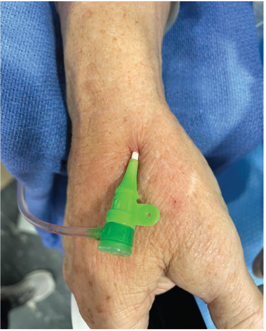
The patient’s forearm and hand are prepared as usual, and the patient’s hand is in a handshake position. The access is obtained beyond the snuff box in the first interphalangeal space (Figure 1). Most of the access, at our institution, is based on palpation, with rare occasions of ultrasound-assisted access. During ultrasound evaluation, the artery is found just under the cephalic vein with two radial veins accompanying it on either side.
Our practice has been double puncture of the vessel with placement of a hydrophilic wire, over which a Terumo 6 French Slender sheath is placed. The radial cocktail (4K units of heparin, 200 units NTG, and 2 mg verapamil) is injected as usual and flushed. A Tegaderm (3M) with a central square window is placed over the sheath to anchor it in situ. Six French diagnostic and interventional catheters are used for the procedure.
Post procedure, the sheath is pulled before the patient leaves the catheterization laboratory, for both diagnostic and interventional procedures. A 2x2 gauze is placed on the access site. With firm finger pressure on the gauze, a Coban bandage (3M) is applied on the access site. Coban is placed in a circumferential fashion around the wrist for 2 to 3 rounds, followed by a figure-of-eight bandage, made by bringing the Coban over the first interphalangeal space,
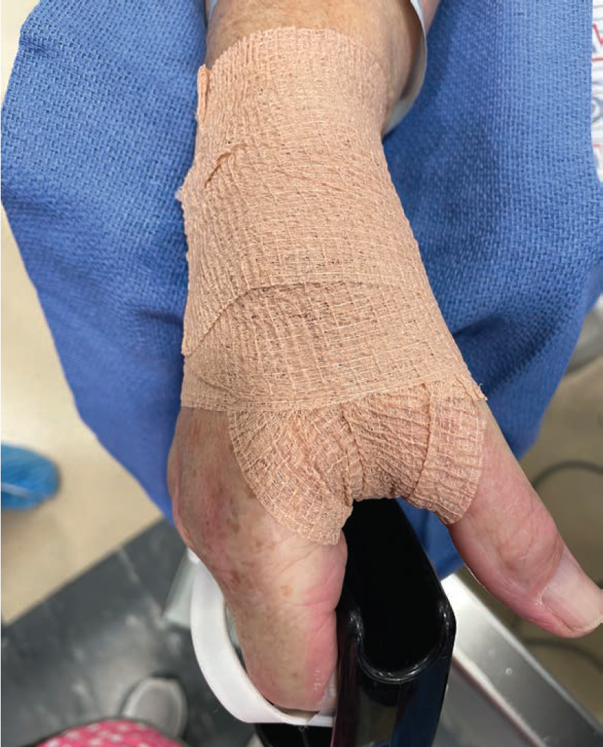
with a circumferential roll to end it all (Figure 2). This bandage is kept on for 1 hour post diagnostic procedure and 2 hours post intervention. Throughout this period, a pulse oximeter is hooked up to one of the fingers of the bandaged hand to ensure appropriate distal flow. By protocol, the figure-of-eight bandage through the first interphalangeal space is unwrapped at 30 minutes and 60 minutes post diagnostic and interventional procedures, respectively, and the second layer removed 60 minutes and 120 minutes later (also respectively). A small Tegaderm is then placed over a folded gauze. The patient is watched for any bleeding over the next 1 to 3 hours. If any rebleed is noted, manual pressure with reapplication of the Coban bandage for an additional 30 to 60 minutes occurs.
Impact
Our experience has been that the distal dorsal access site is very conducive to patient comfort, with a neutral position of the hand during the procedure. We have observed reduced events of vasospasm and pain compared to standard radial access, with definitive hemostasis prior to leaving the laboratory. The Coban bandage has a feel of a boxer bandage rather than a high-pressure radial hemostasis device post procedure for the patient. From an operator standpoint, the position of the arm is very helpful during the procedure with less patient movement, and the reduced occurrence of vasospasm we have observed has been notable and very helpful.
Post-procedure control on the access site with hemostasis is comforting. If any bleeding is noted post bandage removal, firm manual pressure with reapplication of the Coban bandage is quite a simple process that does not need any special training. The nursing staff seem to prefer the distal dorsal access with bandage over a radial hemostasis device follow-up post procedure due to its simplicity and efficacy.
Post access site care of a cardiac catheterization patient is a significant issue in smaller community hospitals. In the present hospital environment of constant staff flux, the issues of training staff and retaining trained staff over any significant period of time are real and vexing for all parties involved. The distinct advantage of the distal dorsal approach is very helpful in these situations, with lower stress for the operating physicians as well.
An additional advantage of the distal dorsal radial access site has been noted in radiocephalic fistula interventions. The additional working length available with this access means interventions at the anastomotic site and the proximal limb of the arteriovenous fistula are feasible and more convenient.
Final Advice
A few notes of caution to the operator. It is critical to make sure the hydrophilic guide wire, immediately after vessel access, is moving freely in the north-south direction prior to placement of the sheath. Occasional tactile feel of the wire entering aberrant branches, side branches, and/or the dorsal arch does occur. It is felt as a resistance to wire movement and difficulty in wire torque. Under such situations, the wire is withdrawn and redirected with constant rotational movement, making sure the north-south movement of the wire is free. The access needle angio-catheter is removed only after confirming this free movement, followed by placement of the 6 French sheath over this wire. Also, in patients with bradycardic rhythm, both the introduction and withdrawal of the needle sheath/catheter need to be done very slowly. Finally, this approach may be an impractical option in very tall individuals, due to the limitations in length of available diagnostic and interventional catheters.
Notes From a Community Hospital Operator: How It All Came About
Ananth N. Kumar, MD, FACC
Life’s serendipity and circumstances have made me a firm believer in the value of a subspecialist in a community hospital setting. This symbiotic relationship can lead to immense professional satisfaction for the provider and high quality, valuable, local personable care for the community.
I have been extraordinarily fortunate to have had opportunities to start two successful “primary angioplasty without surgical backup” programs in the community hospital setting over the last 12 years. Being a solo provider for the majority of the time, a constant effort to bring the latest and best practices to our little paradise became the modus operandi. Thanks to highly dedicated, loyal, amazing staff, we have been successful in making these programs an asset to the community with high quality standards, including a median door-to-balloon (D2B) time consistently 15 to 20 minutes less than the top 90th percentile of hospitals nationwide, for over five years.
In this setting, major medical conferences became tools of self-assessment and improvement. At one of these conferences, in 2017, I attended a dedicated talk on radial access. The esteemed presenter (unfortunate that I do not recollect the name) concluded his talk with the remark, “You should try distal radial access — you will never go radial again.” That statement stirred my interest and I promptly started implementing distal dorsal radial access immediately after. Subsequently published journal literature kept affirming the path that we were on. True to the presenter’s words, we were a majority distal dorsal radial lab within a few months. The learning curve was not steep and the benefits were obvious. I continue to be grateful for his talk so many years ago that spurred us to make such a positive change in the overall cath lab experience for all parties involved.
References
1. Fazel R, Rao SV, Cohen DJ, et al. Radial vs femoral access for percutaneous coronary intervention: temporal trends and outcomes in the USA. Eur Heart J. 2025 Jul 4: ehaf426. doi:10.1093/eurheartj/ehaf426
2. Rashid M, Kwok CS, Pancholy S, et al. Radial artery occlusion after transradial interventions: a systematic review and meta-analysis. J Am Heart Assoc. 2016 Jan 25; 5(1): e002686. doi:10.1161/JAHA.115.002686
3. Sampath-Kumar R, Mahmud E, Korkmaz K, et al. Patient characteristics and outcomes of radial to femoral access-site crossover. J Soc Cardiovasc Angiogr Interv. 2025 Jan 21; 4(1):102450. doi:10.1016/j.jscai.2024.102450
4. Aoi S, Htun WW, Freeo S, et al. Distal transradial artery access in the anatomical snuffbox for coronary angiography as an alternative access site for faster hemostasis. Catheter Cardiovasc Interv. 2019 Nov 1; 94(5): 651-657. doi:10.1002/ccd.28155
5. Mahmoud MAT, Hamam NG, Ghanm TIE, et al. Comparing distal and proximal radial access for percutaneous coronary intervention and angiography: a comprehensive meta-analysis and systematic review of randomized controlled trials. Coron Artery Dis. 2025 Sep 1; 36(6): e1-e11. doi:10.1097/MCA.0000000000001489
Find More:
Renal Denervation Topic Center
Cardiovascular Ambulatory Surgery Centers (ASCs) Topic Center
Grand Rounds With Morton Kern, MD
Peripheral Artery Disease Topic Center


