


Lode Ergometer Being Used by UF Health to Help Diagnose Hypertrophic Cardiomyopathy
By Bill Levesque
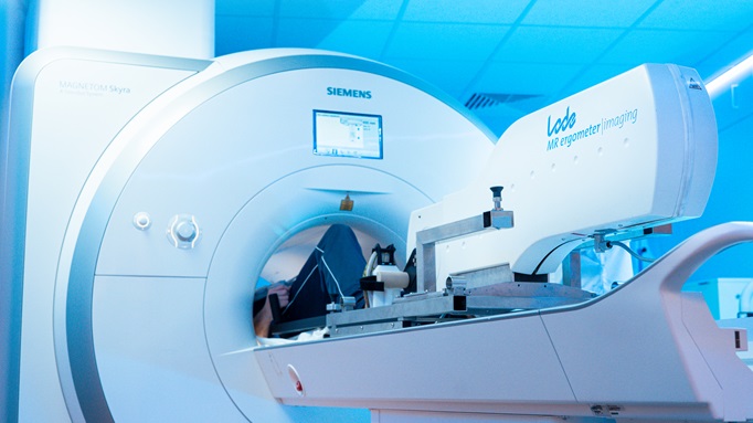
GAINESVILLE, Fla. — The device resembles an odd stationary bicycle without a seat or handlebars. Anyone who sees it at University of Florida Health would quickly realize there’s only one way to pedal — lying supine inside an MRI machine.
UF Health cardiologists are using a new diagnostic tool called a pedal ergometer that will help doctors more quickly identify a notoriously hard-to-detect heart condition, hypertrophic cardiomyopathy, or HCM, so that patients can more rapidly begin life-changing treatments.
The inherited disease, an abnormal thickening of heart muscle, makes it more difficult for the heart to pump blood and affects 1 in 200 people. HCM can cause fatigue, chest pain, shortness of breath, arrhythmias and, in the worst cases, heart failure and death.
Using a nearly $400,000 grant from the pharmaceutical company Bristol Myers Squibb, UF Health recently obtained the specialized, MRI-compliant ergometer to allow stress tests to detect HCM. (An ergometer measures the work done by muscles.)
Mustafa M. Ahmed, M.D., a professor in the UF College of Medicine’s division of cardiovascular medicine and its heart failure section chief, said UF Health is likely the first health system in the region to use such a device.
“It’s very novel,” Ahmed said. “It allows us to differentiate some other very nuanced diagnoses in addition to hypertrophic cardiomyopathy. That really enhances our ability to be at the leading edge of imaging diagnostics for cardiac disease processes.”
The ergometer, supported by a table, attaches to the front of the opening where a patient lies in the MRI machine. The patient pedals for several minutes during the tests.
The device contains no metallic components, so it is not affected by the MRI’s powerful magnets.
The telltale signs of HCM are more apparent during exertion because the condition worsens as the heart works harder.
“As you are obtaining cardiac MRI images, you can see how the heart is responding to exercise throughout that imaging sequence,” Ahmed said.
HCM can be difficult to detect because patients often have either no or mild symptoms. And those symptoms can mimic other conditions, such as asthma, overexertion or fatigue. Only an estimated 15% of affected patients know they have the condition.
“It can be a tricky diagnosis to make, and oftentimes we make this diagnosis with echocardiography,” Ahmed said. “The MRI allows you to characterize the heart muscle tissue itself, and that sometimes has implications regarding prognosis or regarding the need for other types of therapies or things like defibrillators or specialized medical therapy.”
UF Health cardiologists can then better distinguish HCM from conditions with similar features, such as “athlete’s heart,” which causes changes to the heart due to frequent strenuous exercise.
HCM often has a debilitating impact on a patient’s quality of life. Treatment has advanced in recent years, including the development of cardiac myosin inhibitors, a class of drugs that targets the disease at the molecular level.
The MRI-compliant bicycle is part of a broader quality improvement initiative at UF Health to streamline and improve diagnostic capabilities to treat the disease.
The goal is to have UF Health’s HCM practice become a Hypertrophic Cardiomyopathy Association “Center of Excellence.”
The division of cardiovascular medicine is collaborating on the project with the UF College of Medicine’s Department of Radiology, UF Continuing Medical Education and the PeerView Institute for Medical Education.
Find More:
Renal Denervation Topic Center
Cardiovascular Ambulatory Surgery Centers (ASCs) Topic Center
Grand Rounds With Morton Kern, MD
Peripheral Artery Disease Topic Center
Podcasts: Cath Lab Conversations


