Hard to Crack: The Persistent Challenge of Peripheral Arterial Calcium
© 2025 HMP Global. All Rights Reserved.
Any views and opinions expressed are those of the author(s) and/or participants and do not necessarily reflect the views, policy, or position of Vascular Disease Management or HMP Global, their employees, and affiliates.
VASCULAR DISEASE MANAGEMENT. 2025;22(12):E115-E117

Cardiovascular Institute of the South, Houma, Louisiana
Vascular calcification in peripheral arterial disease (PAD) is commonly thought of as a product of age. While it does constitute a part of the normal aging process, conditions such as diabetes mellitus (DM), chronic kidney disease (CKD), tobacco use, and hypertension play a large role in pathologic calcium deposition, even in younger individuals.1 The mechanism of intimal and medial wall vascular calcification is central to smooth muscle cells. In intimal calcification, lipid deposits lead to macrophage invasion, activation, and subsequent smooth muscle proliferation. This then forms atherosclerotic plaques. In medial calcification, smooth muscle cells differentiate into osteoblast-like cells and directly deposit calcium into the vessel wall. The latter, previously referred to as Mönckeberg medial calcification, was historically thought to be benign in nature, but it also contributes to cardiovascular morbidity and mortality and additionally provides significant difficulty and challenges with peripheral intervention.2
Several interventional treatment challenges are set forth by vascular calcification. These include increased rates of restenosis after intervention, vessel dissection and perforation, inadequate stent expansion, and distal embolization.3 Moreover, commonly used treatment modalities such as drug-coated balloons (DCB) and drug-eluting stents are rendered less effective by the presence of vessel calcification.4,5 Technological advancements in the peripheral space have helped to address these issues; however, dense calcium continues to be the single-biggest obstacle for the interventionalist when treating PAD.
The cases presented by Finn et al in “Dealing With Dense Calcium” illustrate 3 interesting points regarding dense calcium and intervention. First, several devices exist that provide varying methods of calcium modification and debulking after wire crossing of the lesion. These devices have fundamental differences that dictate how the calcium will be modified; one may provide case-specific advantages over another, depending on the clinical context. Second, newer tools exist that can aid in balloon or wire-uncrossable lesions in the setting of dense calcium when other tools have failed. Finally and possibly most importantly, the authors show how critical imaging, access site, and use of alternative access can be in dealing with dense calcium—a strategy that can be employed even in labs without access to some of these newer tools.
Case 1 illustrates one of the tools currently available in many labs for modification of calcium—LASER atherectomy. There are currently 2 excimer laser atherectomy systems in routine use in the United States for the treatment of PAD. These include the Philips 308 nanometer system and the Auryon 355 nanometer system (Angiodynamics). Both laser systems facilitate plaque removal by 3 mechanisms: photoablation, sonic pulse wave creation, and cavitation bubble formation. Despite historical anecdotes suggesting laser is effective only in modifying soft plaque or neointimal tissues, the author demonstrates its use in crossing a densely calcified lesion by modifying the proximal lesion cap after multiple failed attempts with standard crossing techniques. The same laser was used to further debulk the body of the lesion. The effectiveness of this device is likely facilitated by the aforementioned sonic wave creation in which each laser pulse helps to shatter hard, calcified areas of the vessel. While effective for intimal calcification involving the vascular lumen, much of the speculation around the efficacy of laser in treating calcific lesions lies in its limited ability to adequately modify medial calcification. To address this, the authors utilized intravascular lithotripsy (IVL) for even further calcium modification prior to the use of a DCB. The series of DISRUPT PAD trials (I and II) clearly demonstrated that IVL was a safe and effective way to dilate calcified lesions with less pressure and fewer dissections than standard percutaneous transluminal angioplasty (PTA).6,7 DISRUPT PAD III demonstrated the superiority of IVL over standard angioplasty prior to DCB treatment or stenting in regard to 1- and 2-year primary patency rates (a secondary endpoint).8 This technology may more effectively treat hard-to-address medial calcific disease than other modalities, making it a unique addition to the interventionalist toolbox.
Case 3 illustrates one of the most recent additions to the IVL family: forward-facing lithotripsy. The Javelin catheter (Shockwave Medical) is a 0.014-inch compatible lithotripsy system that utilizes a forward-facing emitter to deliver sonic shockwaves distal to the tip of the catheter. This can facilitate either balloon or other device passage through a “balloon-uncrossable” lesion. It has even been used in case reports to facilitate wire passage in much the same way as the authors used laser atherectomy in Case 1.
In addition to the modalities listed above, several others have shown promise in calcium modification. The CALCIUM 360 trial evaluated orbital atherectomy (OA) + PTA vs PTA only for treatment of calcified infrapopliteal lesions in patients with chronic limb-threatening ischemia. Results showed significantly lower balloon inflation pressure after OA + PTA as well as significantly higher freedom from major adverse events in the OA + PTA group.9 Observational data utilizing directional atherectomy (DA) in the Total REALITY study showed a significantly lower provisional stent rate when using DA + DCB compared to PTA + DCB despite significantly greater baseline calcification in the DA + DCB group.10 Several specialty balloons also exist with the intention to modify calcium. These include scoring and cutting balloons as well as high-pressure noncompliant balloons. The underlying concept is to create linear fractures in the calcium to assist in lesion expansion and potentially drug absorption. In scoring and cutting balloons, this is done through focusing pressure over a smaller surface area to create controlled plaque incision with less barotrauma to the lesion.
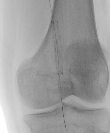
Case 2 focuses on a central but commonly overlooked tenant in dealing with dense calcium—defining the degree of target vessel calcification. It also shows the importance of routine alternative access site selection for difficult-to-treat lesions. Several calcium scoring systems are currently in use, and while many of these have been published in peer-reviewed journals or used in investigational trial core labs (Peripheral Academic Research Consortium scale and Peripheral Arterial Calcium Scoring Scale), it is important to note that none of them have been validated by hard endpoints. In general, they rely on either angiographic or intravascular imaging parameters such as calcification on both sides of the vessel or circumferential degree of calcium, respectively. The authors note that prior to intervention, a computed tomography (CT) was obtained, demonstrating severe calcific popliteal and infrapopliteal disease. Not only does this aid in calcium detection and distribution, but it also allows assessment of potential alternative access sites. The authors further state that extravascular ultrasound was used to first obtain distal access in the calcified posterior tibial artery and subsequently to navigate the dense calcium present in the vessel. Next, intravascular ultrasound was used to assess the degree of calcification in this artery to choose a treatment modality and to appropriately size the vessel. Despite its overwhelmingly supportive data and clinical utility, routine use of intravascular imaging in clinical practice is still lacking as it is not uniformly reimbursed. It is often seen as an added expense to already high-dollar procedures. The authors were able to use this modality as well as extravascular ultrasound and pre-procedure CT to avoid potential antegrade wiring or treatment complications and likely decrease overall procedure time.
This case series by Finn et al highlights several problems with dense calcium from an intervention standpoint. It provides several real-world examples of how advances in device technology and procedure setup can be used to safely and effectively navigate calcified lesions that previously would have been deemed “untreatable.” Unfortunately, densely calcified lesions will become an even greater problem in the future with the general population having increased life expectancy, increased incidence of DM, and more CKD. Despite procedural advances, interventions such as smoking cessation, exercise programs, and strict blood glucose and blood pressure control will remain the foundation of preventive treatment. n
References
1. Lee SJ, Lee I, Jeon J. Vascular calcification—new insights into its mechanism. Int J Mol Sci. 2020;21(8):2685. doi:10.3390/ijms21082685
2. Van den Bergh G, Opdebeeck B, D’Haese PC, Verhulst A. The vicious cycle of arterial stiffness and arterial media calcification. Trends Mol Med. 2019;25(12):1133-1146. doi:10.1016/j.molmed.2019.08.006
3. Rocha-Singh KJ, Zeller T, Jaff MR. Peripheral arterial calcification: prevalence, mechanism, detection, and clinical implications. Catheter Cardiovasc Interv. 2014;83(6):E212-E220. doi:10.1002/ccd.25387
4. Kawaguchi R, Tsurugaya H, Hoshizaki H, Toyama T, Oshima S, Taniguchi K. Impact of lesion calcification on clinical and angiographic outcome after sirolimus-eluting stent implantation in real-world patients. Cardiovasc Revasc Med. 2008;9(1):2-8. doi:10.1016/j.carrev.2007.07.004
5. Fanelli F, Cannavale A, Gazzetti M, et al. Calcium burden assessment and impact on drug-eluting balloons in peripheral arterial disease. Cardiovasc Intervent Radiol. 2014;37(4):898-907. doi:10.1007/s00270-014-0904-3
6. Brodmann M, Werner M, Brinton TJ, et al. Safety and performance of lithoplasty for treatment of calcified peripheral artery lesions. J Am Coll Cardiol. 2017;70(7):908-910. doi:10.1016/j.jacc.2017.06.022
7. Brodmann M, Werner M, Holden A, et al. Primary outcomes and mechanism of action of intravascular lithotripsy in calcified, femoropopliteal lesions: results of Disrupt PAD II. Catheter Cardiovasc Interv. 2019;93(2):335-342. doi:10.1002/ccd.27943
8. Tepe G, Brodmann M, Bachinsky W, et al. Intravascular lithotripsy for peripheral artery calcification: mid-term outcomes from the randomized Disrupt PAD III trial. J Soc Cardiovasc Angiogr Interv. 2022;1(4):100341. doi:10.1016/j.jscai.2022.100341
9. Shammas NW, Lam R, Mustapha J, et al. Comparison of orbital atherectomy plus balloon angioplasty vs. balloon angioplasty alone in patients with critical limb ischemia: results of the CALCIUM 360 randomized pilot trial. J Endovasc Ther. 2012;19(4):480-488. doi:10.1583/JEVT-12-3815MR.1
10. Medtronic presents studies on utility of atherectomy to treat PAD. Endovascular Today. Accessed December 14, 2025. https://evtoday.com/news/medtronic-presents-studies-on-utility-of-atherectomy-to-treat-pad