Ultrasound as a Transformative Tool in Dermatology: From Cosmetic Imaging to Hidradenitis Suppurativa Assessment and Emerging Therapies
Dermatologic ultrasound is increasingly recognized as a practical, point-of-care imaging tool with broad clinical utility across both medical and cosmetic dermatology. At the 2026 Masterclasses in Dermatology Annual Meeting, Jane Yoo, MD, MPP (janeyoomd.com), and Alice Bendix Gottlieb, MD, PhD, reviewed the principles and applications of handheld ultrasound devices, sonographic assessment of hidradenitis suppurativa (HS), aesthetic procedure guidance, and the expanding landscape of HS therapeutics.
Dr Yoo framed dermatologic ultrasound as the "fourth eye," an extension of clinical visualization beyond the naked eye and dermoscopy. Handheld devices connect wirelessly to smartphones and emit high-frequency acoustic waves that reflect off skin structures based on tissue density. Denser structures appear brighter (hyperechoic) while fluid-filled structures appear darker (hypoechoic or anechoic), forming the interpretive foundation of dermatologic ultrasound. These properties allow clinicians to characterize skin lesions, assess inflammatory conditions, identify filler material type and placement, and map vascular anatomy prior to cosmetic injections.
In the aesthetic setting, ultrasound enables preprocedural mapping of facial danger zones, including the glabella, temple, nasolabial fold, perioral region, and infraorbital area. Real-time guidance supports precise placement of fillers and neuromodulators and allows targeted dissolution of excess or misplaced filler with hyaluronidase. Each filler material carries a distinct sonographic signature. Hyaluronic acid appears as well-defined anechoic deposits, calcium hydroxyapatite produces hyperechoic bands with posterior acoustic shadowing, and silicone generates a characteristic "snowstorm" reverberation pattern. In vascular adverse events, Doppler imaging can confirm arterial compromise from intravascular filler and guide hyaluronidase delivery directly to the obstructing deposit, with documented recovery of arterial flow following treatment.
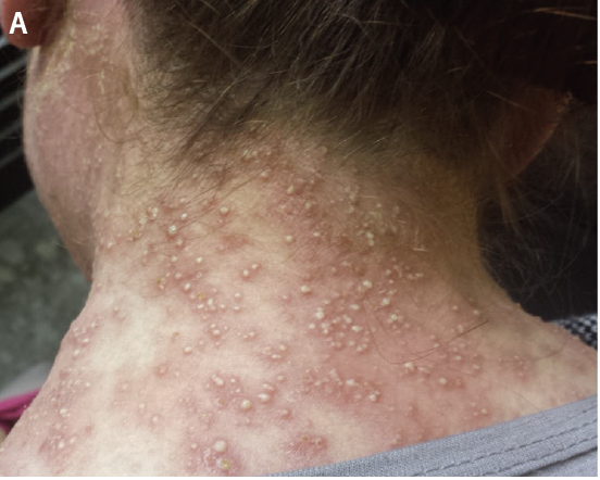
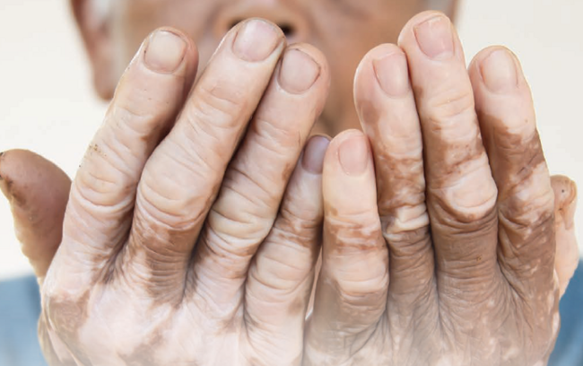
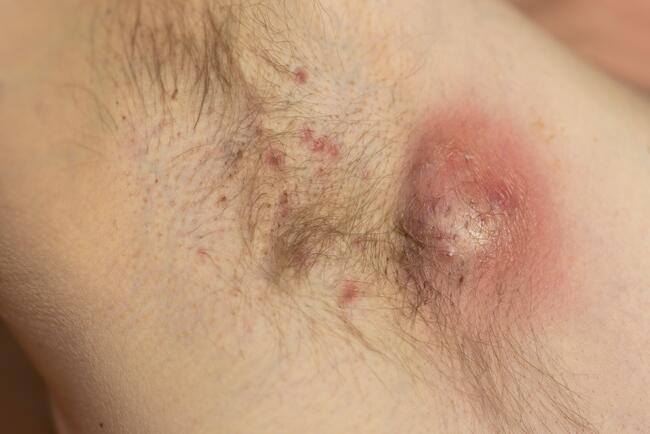
In HS, ultrasound detects tunnels, abscesses, and subclinical scarring before these features become clinically apparent. Sonographic criteria for HS include widening of hair follicles, dermal thickening, pseudocystic nodules, fluid collections, and fistulous tracts, with at least 3 required for diagnosis. Two pathognomonic signs have been characterized: the donor sign, representing ballooned hair follicles within a fluid collection or fistulous tract, and the bridge sign, a hypoechoic band connecting 2 or more follicles. Dr Gottlieb emphasized that the ability to identify subclinical disease supports earlier intervention to prevent scarring, chronic draining tunnels, disability, and diminished quality of life.
Dr Gottlieb reviewed the current HS treatment landscape. Three biologics are US Food and Drug Administration (FDA) approved: adalimumab (TNF-α inhibitor), approved for adults and adolescents age 12 years and older; secukinumab (IL-17A inhibitor); and bimekizumab (IL-17A/F inhibitor). Infliximab remains widely used off label as the only truly weight-based intravenous biologic option. Several agents are in advanced development. Sonelokimab, an IL-17A/F nanobody in phase 3, achieved HiScr75 in 34% of patients vs 17.5 to 24.9% for placebo. Povorcitinib, a JAK inhibitor in the phase 3 STOP-HS program, achieved HiScr50 in approximately 40% of patients vs 29.7% for placebo, with a second pivotal trial yielding similar results. Lutikizumab, a dual IL-1α/IL-1β monoclonal antibody, achieved HiScr50 in 66% of patients in a single-arm phase 2 study. Conventional therapies, including antibiotics, spironolactone, colchicine, dapsone, and surgery, continue to play a role in management, although many lack FDA approval specifically for HS.
As the dermatologic toolkit expands, integration of point-of-care ultrasound alongside targeted systemic therapies represents a meaningful advancement toward earlier diagnosis, more precise treatment, and improved outcomes for patients with HS and those undergoing cosmetic procedures.
Reference
Yoo J, Gottlieb AB. Transforming the dermatologic toolkit with ultrasound: multifunctional applications from cosmetic imaging to HS assessment. Presented at: Masterclasses in Dermatology; February 19–22, 2026; Sarasota, FL.