


Should You Consider Using Bioengineered Alternative Tissues Over Tendon And Bone?
August 2014
Given the inherent obstacles with complex lower extremity wounds, the need for immediate coverage and possible risks with using flaps and pedicles, these authors explore the potential of employing bioengineered alternative tissues in wounds with exposed tendon and bone.
 Complex lower extremity wounds with exposed tendon and bone are a reconstructive challenge. Surgeons have used free tissue transfer as the treatment of choice in order to achieve adequate soft tissue coverage. In certain settings, however, free tissue transfer or local flap coverage is associated with increased morbidity and mortality, and may not be the best option.
Unfortunately, in these situations, amputation may be the treatment of choice or the end result.1-6 The development of bioengineered alternative tissues (BATs) has led to new methods for reconstruction of these complex wounds. When free tissue transfer is not an option, surgeons have turned to the use of BAT in combination with split-thickness skin grafting (STSG) for adequate wound coverage.2,5 Specifically, one can use dermoconductive BATs (those with no cellular components) to serve as scaffolds for cellular migration for deep tissue coverage and neodermis production.6-8 Due to potential complications associated with complex microsurgical procedures, this method of wound coverage is becoming increasingly popular.
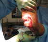
As soon as a wound on the lower extremity presents with exposed tendon or bone, the need for quick and effective coverage becomes a more daunting situation. Trauma such as severe crush injuries, pressure ulcers, chronic diabetic ulcers, venous or arterial ulcers, and surgical wound complications can all lead to deep exposure of these structures. Without the exposure of such structures, typical management would be debridement, local wound care with dressings and offloading. Due to the biology of tendons and bone, the need for immediate and definitive coverage is key.3,5,9
The main issue with deep, complex wounds of the lower extremity is that the wound itself does not provide the adequate blood supply to keep a skin graft alive. The lower extremity also has limited options for local tissue transfer. Surgeons have accordingly turned to free tissue transfer. In many patients the length and intricacy of these procedures is contraindicated and can lead to significant complications. Contraindications for free flaps include significant comorbidities, hemodynamic instability, age, inadequate blood flow and infection. Things to consider prior to surgery are the length of the procedure, donor site morbidity, the need for close monitoring and potential failure.3,4
Techniques using flaps and pedicles are associated with a failure rate of 12 percent and an amputation rate of 18 percent.3,10-13 There has also been an estimated length of stay of about 76 hospital days with an average of eight surgeries.3,10-13 It is evident that a simpler method of wound coverage would be more ideal and one should possibly attempt this prior to free tissue transfer in some patients.
Blood vessels and synovial fluid nourish tendons. When tendons lose overlying soft tissue, there is disrupted nourishment and tendons can quickly become desiccated. This causes loss of the tendon fibers as well as loss of proper function of the tendon. Tendons can quickly lose their ability to glide and operate at joints, leading to the need for other surgical interventions such as joint fusions or tendon transfers. In order to avoid these procedures, surgeons aim for quick and definitive coverage.9
Similar to tendons, bones need quick and definitive coverage. The most important reason is to decrease the risk of infection. Osteomyelitis can be a devastating sequela of wounds with exposed bone. Timely soft tissue coverage of bone also increases the potential for healing any fractures that are present.14,15
Complex lower extremity wounds with exposed tendon and bone are a reconstructive challenge. Surgeons have used free tissue transfer as the treatment of choice in order to achieve adequate soft tissue coverage. In certain settings, however, free tissue transfer or local flap coverage is associated with increased morbidity and mortality, and may not be the best option.
Unfortunately, in these situations, amputation may be the treatment of choice or the end result.1-6 The development of bioengineered alternative tissues (BATs) has led to new methods for reconstruction of these complex wounds. When free tissue transfer is not an option, surgeons have turned to the use of BAT in combination with split-thickness skin grafting (STSG) for adequate wound coverage.2,5 Specifically, one can use dermoconductive BATs (those with no cellular components) to serve as scaffolds for cellular migration for deep tissue coverage and neodermis production.6-8 Due to potential complications associated with complex microsurgical procedures, this method of wound coverage is becoming increasingly popular.
As soon as a wound on the lower extremity presents with exposed tendon or bone, the need for quick and effective coverage becomes a more daunting situation. Trauma such as severe crush injuries, pressure ulcers, chronic diabetic ulcers, venous or arterial ulcers, and surgical wound complications can all lead to deep exposure of these structures. Without the exposure of such structures, typical management would be debridement, local wound care with dressings and offloading. Due to the biology of tendons and bone, the need for immediate and definitive coverage is key.3,5,9
The main issue with deep, complex wounds of the lower extremity is that the wound itself does not provide the adequate blood supply to keep a skin graft alive. The lower extremity also has limited options for local tissue transfer. Surgeons have accordingly turned to free tissue transfer. In many patients the length and intricacy of these procedures is contraindicated and can lead to significant complications. Contraindications for free flaps include significant comorbidities, hemodynamic instability, age, inadequate blood flow and infection. Things to consider prior to surgery are the length of the procedure, donor site morbidity, the need for close monitoring and potential failure.3,4
Techniques using flaps and pedicles are associated with a failure rate of 12 percent and an amputation rate of 18 percent.3,10-13 There has also been an estimated length of stay of about 76 hospital days with an average of eight surgeries.3,10-13 It is evident that a simpler method of wound coverage would be more ideal and one should possibly attempt this prior to free tissue transfer in some patients.
Blood vessels and synovial fluid nourish tendons. When tendons lose overlying soft tissue, there is disrupted nourishment and tendons can quickly become desiccated. This causes loss of the tendon fibers as well as loss of proper function of the tendon. Tendons can quickly lose their ability to glide and operate at joints, leading to the need for other surgical interventions such as joint fusions or tendon transfers. In order to avoid these procedures, surgeons aim for quick and definitive coverage.9
Similar to tendons, bones need quick and definitive coverage. The most important reason is to decrease the risk of infection. Osteomyelitis can be a devastating sequela of wounds with exposed bone. Timely soft tissue coverage of bone also increases the potential for healing any fractures that are present.14,15
 To bypass the need for technically demanding procedures, physicians have employed bioengineered alternative tissues as a much simpler and less demanding alternative. Researchers have recently begun to study Integra (Integra Life Sciences) and Graftjacket (KCI) for this use. Minimal data is available but there are several small case studies that evaluate the use and outcomes of these products over tendon and bone. Both of these products provide a collagen scaffold that allows for vascular in-growth.1,16,17 The most frequently encountered complications with such grafts are seroma and hematoma. In order to decrease the incidence of these complications, surgeons have combined application of the graft with negative pressure wound therapy (NPWT).1,12,18
 In 2007, Helgeson and colleagues from the Walter Reed Army Medical Center studied their experience with Integra on combat-related wounds.17 They looked at 16 wounds caused by blast-related accidents. Of the 16 wounds, 11 had exposed tendon and absence of paratenon, and the remaining five wounds had exposed bone without periosteum. The researchers performed an average of 8.5 irrigations and debridements per wound prior to application of Integra. Some wounds required “stacking” of the graft in order to fill a potential void. Physicians applied VAC dressings to all wounds until the neodermis formed and did dressing changes every three to four days. The study researchers subsequently applied a STSG an average of 19 days after application of the Integra graft.
There was definitive coverage in 13 out of 16 (81 percent) of the original wounds after the initial Integra application.17 Of the three wounds that failed, all had exposed bone. Two went on to heal after a second Integra application. The third wound was located at the hip and healed after several months of VAC therapy. The authors stated that in their particular patient population, flap coverage is rather challenging due to such large zones of injury and inflammation, and Integra with VAC and STSG is a successful alternative.
There have been similar results in several other small studies. Menn and coworkers saw stable wound healing in four elderly patients with multiple medical problems.1 Three had STSG after an average of 29 days and one patient healed by secondary intention. Lee and coworkers reported the use of Integra and STSG over burn injuries with exposed tendon or bone in seven patients.5 They saw complete wound healing of all seven patients with only two patients requiring a second application of Integra. Another study by Shores and coworkers reported the results of using Integra in 42 patients with exposed tendons from trauma, excision of cancer tumor wounds or chronic wounds.23 They saw a 92.5 percent take rate after STSG and a 91.2 percent attainment of range of motion in comparison to the contralateral side.
Fraccalvieri, Clerici and their respective colleagues looked at calcaneal osteomyelitis and acute foot infections that resulted in exposed tendon or bone.24,25 They saw 100 percent and 86.7 percent healing rates respectively with the use of Integra on their vasculopathic patients and patients with diabetes. There is minimal evidence in regard to the use of Integra over chronically exposed deep structures.
In 2007, Helgeson and colleagues from the Walter Reed Army Medical Center studied their experience with Integra on combat-related wounds.17 They looked at 16 wounds caused by blast-related accidents. Of the 16 wounds, 11 had exposed tendon and absence of paratenon, and the remaining five wounds had exposed bone without periosteum. The researchers performed an average of 8.5 irrigations and debridements per wound prior to application of Integra. Some wounds required “stacking” of the graft in order to fill a potential void. Physicians applied VAC dressings to all wounds until the neodermis formed and did dressing changes every three to four days. The study researchers subsequently applied a STSG an average of 19 days after application of the Integra graft.
There was definitive coverage in 13 out of 16 (81 percent) of the original wounds after the initial Integra application.17 Of the three wounds that failed, all had exposed bone. Two went on to heal after a second Integra application. The third wound was located at the hip and healed after several months of VAC therapy. The authors stated that in their particular patient population, flap coverage is rather challenging due to such large zones of injury and inflammation, and Integra with VAC and STSG is a successful alternative.
There have been similar results in several other small studies. Menn and coworkers saw stable wound healing in four elderly patients with multiple medical problems.1 Three had STSG after an average of 29 days and one patient healed by secondary intention. Lee and coworkers reported the use of Integra and STSG over burn injuries with exposed tendon or bone in seven patients.5 They saw complete wound healing of all seven patients with only two patients requiring a second application of Integra. Another study by Shores and coworkers reported the results of using Integra in 42 patients with exposed tendons from trauma, excision of cancer tumor wounds or chronic wounds.23 They saw a 92.5 percent take rate after STSG and a 91.2 percent attainment of range of motion in comparison to the contralateral side.
Fraccalvieri, Clerici and their respective colleagues looked at calcaneal osteomyelitis and acute foot infections that resulted in exposed tendon or bone.24,25 They saw 100 percent and 86.7 percent healing rates respectively with the use of Integra on their vasculopathic patients and patients with diabetes. There is minimal evidence in regard to the use of Integra over chronically exposed deep structures.
 A second study in 2009 looked at seven wounds in patients 2 to 12 years old that were not amenable to primary or secondary wound closure.14 The wounds included were three open tibia fractures, three foot or ankle degloving injuries, and one open ankle fracture. All seven wounds healed without any amputations or need for more complicated flap coverage. Only two patients developed a complication at the Integra or STSG site. One patient had a 1 cm area of delayed healing treated with dressing changes and researchers opted for debridement and secondary closure for the second non-healing wound.
A third study looked at eight patients with an average age of 8.8 years.9 There were seven trauma-related lower extremity wounds with tendon exposed in four patients and bone exposed in six patients. The study authors used Integra, NPWT and skin grafting for all of the wounds. It took on average of 65 days for total wound coverage to occur. There was 100 percent take of the collagen scaffold in six out of the seven lower extremity wounds. The patient with loss of the graft had exposed bone on the lower leg and went on to wound closure with granulation tissue.
A second study in 2009 looked at seven wounds in patients 2 to 12 years old that were not amenable to primary or secondary wound closure.14 The wounds included were three open tibia fractures, three foot or ankle degloving injuries, and one open ankle fracture. All seven wounds healed without any amputations or need for more complicated flap coverage. Only two patients developed a complication at the Integra or STSG site. One patient had a 1 cm area of delayed healing treated with dressing changes and researchers opted for debridement and secondary closure for the second non-healing wound.
A third study looked at eight patients with an average age of 8.8 years.9 There were seven trauma-related lower extremity wounds with tendon exposed in four patients and bone exposed in six patients. The study authors used Integra, NPWT and skin grafting for all of the wounds. It took on average of 65 days for total wound coverage to occur. There was 100 percent take of the collagen scaffold in six out of the seven lower extremity wounds. The patient with loss of the graft had exposed bone on the lower leg and went on to wound closure with granulation tissue.
 Overall, the use of Integra in the challenging pediatric population has negated the need for more complex reconstructive surgeries and patients have tolerated it well in combination with NPWT.
 The third Graftjacket study was a 16-week prospective, randomized, control trial that assessed the use of Graftjacket on full-thickness diabetic wounds in the lower extremity in comparison to sharp debridement alone.28 All wounds were grade 2 according to the Wagner ulcer classification, meaning they extended to tendon, bone or capsule. There were 28 patients with 14 in each treatment group. By week 16, 12 of the 14 patients treated with Graftjacket healed with complete epithelialization. In the same timeframe, only four of the control group wounds healed. There was a statistically significant difference in the rate of wound healing with Graftjacket in comparison to debridement alone.
 Complex lower extremity wounds with exposed tendon and bone are a reconstructive challenge. Surgeons have used free tissue transfer as the treatment of choice in order to achieve adequate soft tissue coverage. In certain settings, however, free tissue transfer or local flap coverage is associated with increased morbidity and mortality, and may not be the best option.
Unfortunately, in these situations, amputation may be the treatment of choice or the end result.1-6 The development of bioengineered alternative tissues (BATs) has led to new methods for reconstruction of these complex wounds. When free tissue transfer is not an option, surgeons have turned to the use of BAT in combination with split-thickness skin grafting (STSG) for adequate wound coverage.2,5 Specifically, one can use dermoconductive BATs (those with no cellular components) to serve as scaffolds for cellular migration for deep tissue coverage and neodermis production.6-8 Due to potential complications associated with complex microsurgical procedures, this method of wound coverage is becoming increasingly popular.
As soon as a wound on the lower extremity presents with exposed tendon or bone, the need for quick and effective coverage becomes a more daunting situation. Trauma such as severe crush injuries, pressure ulcers, chronic diabetic ulcers, venous or arterial ulcers, and surgical wound complications can all lead to deep exposure of these structures. Without the exposure of such structures, typical management would be debridement, local wound care with dressings and offloading. Due to the biology of tendons and bone, the need for immediate and definitive coverage is key.3,5,9
The main issue with deep, complex wounds of the lower extremity is that the wound itself does not provide the adequate blood supply to keep a skin graft alive. The lower extremity also has limited options for local tissue transfer. Surgeons have accordingly turned to free tissue transfer. In many patients the length and intricacy of these procedures is contraindicated and can lead to significant complications. Contraindications for free flaps include significant comorbidities, hemodynamic instability, age, inadequate blood flow and infection. Things to consider prior to surgery are the length of the procedure, donor site morbidity, the need for close monitoring and potential failure.3,4
Techniques using flaps and pedicles are associated with a failure rate of 12 percent and an amputation rate of 18 percent.3,10-13 There has also been an estimated length of stay of about 76 hospital days with an average of eight surgeries.3,10-13 It is evident that a simpler method of wound coverage would be more ideal and one should possibly attempt this prior to free tissue transfer in some patients.
Blood vessels and synovial fluid nourish tendons. When tendons lose overlying soft tissue, there is disrupted nourishment and tendons can quickly become desiccated. This causes loss of the tendon fibers as well as loss of proper function of the tendon. Tendons can quickly lose their ability to glide and operate at joints, leading to the need for other surgical interventions such as joint fusions or tendon transfers. In order to avoid these procedures, surgeons aim for quick and definitive coverage.9
Similar to tendons, bones need quick and definitive coverage. The most important reason is to decrease the risk of infection. Osteomyelitis can be a devastating sequela of wounds with exposed bone. Timely soft tissue coverage of bone also increases the potential for healing any fractures that are present.14,15
Complex lower extremity wounds with exposed tendon and bone are a reconstructive challenge. Surgeons have used free tissue transfer as the treatment of choice in order to achieve adequate soft tissue coverage. In certain settings, however, free tissue transfer or local flap coverage is associated with increased morbidity and mortality, and may not be the best option.
Unfortunately, in these situations, amputation may be the treatment of choice or the end result.1-6 The development of bioengineered alternative tissues (BATs) has led to new methods for reconstruction of these complex wounds. When free tissue transfer is not an option, surgeons have turned to the use of BAT in combination with split-thickness skin grafting (STSG) for adequate wound coverage.2,5 Specifically, one can use dermoconductive BATs (those with no cellular components) to serve as scaffolds for cellular migration for deep tissue coverage and neodermis production.6-8 Due to potential complications associated with complex microsurgical procedures, this method of wound coverage is becoming increasingly popular.
As soon as a wound on the lower extremity presents with exposed tendon or bone, the need for quick and effective coverage becomes a more daunting situation. Trauma such as severe crush injuries, pressure ulcers, chronic diabetic ulcers, venous or arterial ulcers, and surgical wound complications can all lead to deep exposure of these structures. Without the exposure of such structures, typical management would be debridement, local wound care with dressings and offloading. Due to the biology of tendons and bone, the need for immediate and definitive coverage is key.3,5,9
The main issue with deep, complex wounds of the lower extremity is that the wound itself does not provide the adequate blood supply to keep a skin graft alive. The lower extremity also has limited options for local tissue transfer. Surgeons have accordingly turned to free tissue transfer. In many patients the length and intricacy of these procedures is contraindicated and can lead to significant complications. Contraindications for free flaps include significant comorbidities, hemodynamic instability, age, inadequate blood flow and infection. Things to consider prior to surgery are the length of the procedure, donor site morbidity, the need for close monitoring and potential failure.3,4
Techniques using flaps and pedicles are associated with a failure rate of 12 percent and an amputation rate of 18 percent.3,10-13 There has also been an estimated length of stay of about 76 hospital days with an average of eight surgeries.3,10-13 It is evident that a simpler method of wound coverage would be more ideal and one should possibly attempt this prior to free tissue transfer in some patients.
Blood vessels and synovial fluid nourish tendons. When tendons lose overlying soft tissue, there is disrupted nourishment and tendons can quickly become desiccated. This causes loss of the tendon fibers as well as loss of proper function of the tendon. Tendons can quickly lose their ability to glide and operate at joints, leading to the need for other surgical interventions such as joint fusions or tendon transfers. In order to avoid these procedures, surgeons aim for quick and definitive coverage.9
Similar to tendons, bones need quick and definitive coverage. The most important reason is to decrease the risk of infection. Osteomyelitis can be a devastating sequela of wounds with exposed bone. Timely soft tissue coverage of bone also increases the potential for healing any fractures that are present.14,15
 To bypass the need for technically demanding procedures, physicians have employed bioengineered alternative tissues as a much simpler and less demanding alternative. Researchers have recently begun to study Integra (Integra Life Sciences) and Graftjacket (KCI) for this use. Minimal data is available but there are several small case studies that evaluate the use and outcomes of these products over tendon and bone. Both of these products provide a collagen scaffold that allows for vascular in-growth.1,16,17 The most frequently encountered complications with such grafts are seroma and hematoma. In order to decrease the incidence of these complications, surgeons have combined application of the graft with negative pressure wound therapy (NPWT).1,12,18
Current Insights On The Use Of Integra
Yannas and Burke first developed Integra in the late 1970s and physicians were using it for the treatment of burn wounds by the 1980s.19-21 It is structured to be a bilayer product. The first layer is composed of bovine type I collagen and shark chondroitin-6-sulfate. The second layer is a semi-permeable silicone layer that serves as a temporary epidermis.1 The collagen layer has been developed to allow for maximum cellular in-growth while prohibiting the formation of granulation tissue. Since it is an acellular graft it does not rely on the wound itself for successful take. However, Integra does require vascular ingrowth and population of the matrix with host fibroblasts to create its scaffold effect.1,10,11 Integra becomes incorporated and vascularized by the surrounding wound tissue. It can take up to two to four weeks to vascularize the wound because cells need to grow over the Integra scaffold. This is in contrast to a viable cell-containing graft, which needs revascularization within five to seven days. Surgeons typically employ a staged process in which they employ STSG for final coverage after vascularization of the Integra has occurred.5,19,20 Much of the early research on Integra focused on its use in burn injuries. Molnar and colleagues conducted one of the earlier studies examining the use of Integra in combination with Vacuum Assisted Closure (VAC, KCI) therapy in 2004.22 Their study included eight patients. Five of these patients had exposed bone, tendon or joint, and the remaining three patients had exposed bowel or fascia. The wounds were from trauma, wound dehiscence or after tumor excision. For all the grafts, researchers employed VAC therapy over the Integra. On average, it took 7.25 days for Integra incorporation with a 96 percent take and a 93 percent take of STSG. The authors concluded that the use of VAC therapy with the Integra graft speeds the incorporation of the graft into complicated wounds and is also a good alternative for the reconstruction of such wounds. In 2007, Helgeson and colleagues from the Walter Reed Army Medical Center studied their experience with Integra on combat-related wounds.17 They looked at 16 wounds caused by blast-related accidents. Of the 16 wounds, 11 had exposed tendon and absence of paratenon, and the remaining five wounds had exposed bone without periosteum. The researchers performed an average of 8.5 irrigations and debridements per wound prior to application of Integra. Some wounds required “stacking” of the graft in order to fill a potential void. Physicians applied VAC dressings to all wounds until the neodermis formed and did dressing changes every three to four days. The study researchers subsequently applied a STSG an average of 19 days after application of the Integra graft.
There was definitive coverage in 13 out of 16 (81 percent) of the original wounds after the initial Integra application.17 Of the three wounds that failed, all had exposed bone. Two went on to heal after a second Integra application. The third wound was located at the hip and healed after several months of VAC therapy. The authors stated that in their particular patient population, flap coverage is rather challenging due to such large zones of injury and inflammation, and Integra with VAC and STSG is a successful alternative.
There have been similar results in several other small studies. Menn and coworkers saw stable wound healing in four elderly patients with multiple medical problems.1 Three had STSG after an average of 29 days and one patient healed by secondary intention. Lee and coworkers reported the use of Integra and STSG over burn injuries with exposed tendon or bone in seven patients.5 They saw complete wound healing of all seven patients with only two patients requiring a second application of Integra. Another study by Shores and coworkers reported the results of using Integra in 42 patients with exposed tendons from trauma, excision of cancer tumor wounds or chronic wounds.23 They saw a 92.5 percent take rate after STSG and a 91.2 percent attainment of range of motion in comparison to the contralateral side.
Fraccalvieri, Clerici and their respective colleagues looked at calcaneal osteomyelitis and acute foot infections that resulted in exposed tendon or bone.24,25 They saw 100 percent and 86.7 percent healing rates respectively with the use of Integra on their vasculopathic patients and patients with diabetes. There is minimal evidence in regard to the use of Integra over chronically exposed deep structures.
In 2007, Helgeson and colleagues from the Walter Reed Army Medical Center studied their experience with Integra on combat-related wounds.17 They looked at 16 wounds caused by blast-related accidents. Of the 16 wounds, 11 had exposed tendon and absence of paratenon, and the remaining five wounds had exposed bone without periosteum. The researchers performed an average of 8.5 irrigations and debridements per wound prior to application of Integra. Some wounds required “stacking” of the graft in order to fill a potential void. Physicians applied VAC dressings to all wounds until the neodermis formed and did dressing changes every three to four days. The study researchers subsequently applied a STSG an average of 19 days after application of the Integra graft.
There was definitive coverage in 13 out of 16 (81 percent) of the original wounds after the initial Integra application.17 Of the three wounds that failed, all had exposed bone. Two went on to heal after a second Integra application. The third wound was located at the hip and healed after several months of VAC therapy. The authors stated that in their particular patient population, flap coverage is rather challenging due to such large zones of injury and inflammation, and Integra with VAC and STSG is a successful alternative.
There have been similar results in several other small studies. Menn and coworkers saw stable wound healing in four elderly patients with multiple medical problems.1 Three had STSG after an average of 29 days and one patient healed by secondary intention. Lee and coworkers reported the use of Integra and STSG over burn injuries with exposed tendon or bone in seven patients.5 They saw complete wound healing of all seven patients with only two patients requiring a second application of Integra. Another study by Shores and coworkers reported the results of using Integra in 42 patients with exposed tendons from trauma, excision of cancer tumor wounds or chronic wounds.23 They saw a 92.5 percent take rate after STSG and a 91.2 percent attainment of range of motion in comparison to the contralateral side.
Fraccalvieri, Clerici and their respective colleagues looked at calcaneal osteomyelitis and acute foot infections that resulted in exposed tendon or bone.24,25 They saw 100 percent and 86.7 percent healing rates respectively with the use of Integra on their vasculopathic patients and patients with diabetes. There is minimal evidence in regard to the use of Integra over chronically exposed deep structures.
How Effective Is Integra For Chronic Wounds In The Pediatric Population?
Integra has also been successful in complex wounds in children. Violas and colleagues looked at three extensive lower-extremity trauma wounds that were treated with Integra.18 One patient had an exposed patella and the Integra incorporated well over the bony surface. Even though a second operation was necessary for STSG on these pediatric patients, they tolerated grafting well since it negated the need for daily dressing changes and this success occurred over bone exposure. A second study in 2009 looked at seven wounds in patients 2 to 12 years old that were not amenable to primary or secondary wound closure.14 The wounds included were three open tibia fractures, three foot or ankle degloving injuries, and one open ankle fracture. All seven wounds healed without any amputations or need for more complicated flap coverage. Only two patients developed a complication at the Integra or STSG site. One patient had a 1 cm area of delayed healing treated with dressing changes and researchers opted for debridement and secondary closure for the second non-healing wound.
A third study looked at eight patients with an average age of 8.8 years.9 There were seven trauma-related lower extremity wounds with tendon exposed in four patients and bone exposed in six patients. The study authors used Integra, NPWT and skin grafting for all of the wounds. It took on average of 65 days for total wound coverage to occur. There was 100 percent take of the collagen scaffold in six out of the seven lower extremity wounds. The patient with loss of the graft had exposed bone on the lower leg and went on to wound closure with granulation tissue.
A second study in 2009 looked at seven wounds in patients 2 to 12 years old that were not amenable to primary or secondary wound closure.14 The wounds included were three open tibia fractures, three foot or ankle degloving injuries, and one open ankle fracture. All seven wounds healed without any amputations or need for more complicated flap coverage. Only two patients developed a complication at the Integra or STSG site. One patient had a 1 cm area of delayed healing treated with dressing changes and researchers opted for debridement and secondary closure for the second non-healing wound.
A third study looked at eight patients with an average age of 8.8 years.9 There were seven trauma-related lower extremity wounds with tendon exposed in four patients and bone exposed in six patients. The study authors used Integra, NPWT and skin grafting for all of the wounds. It took on average of 65 days for total wound coverage to occur. There was 100 percent take of the collagen scaffold in six out of the seven lower extremity wounds. The patient with loss of the graft had exposed bone on the lower leg and went on to wound closure with granulation tissue.
 Overall, the use of Integra in the challenging pediatric population has negated the need for more complex reconstructive surgeries and patients have tolerated it well in combination with NPWT.
What The Research Says About Graftjacket
Graftjacket is the second type of collagen scaffold that was originally released for coverage over the periosteum. It is an acellular dermal graft derived from human tissues. Only a few studies have reported its success and, more specifically, its use on deep, complex wounds. Martin and colleagues reported on plantar diabetic foot wounds with a University of Texas grade 2A (non-infected and penetrates to tendon or capsule).26 All 17 patients had surgical debridement and application of Graftjacket. The study authors reported healing in 82.4 percent of wounds at an average of 8.9 weeks. These researchers did not evaluate the use of Graftjacket over exposed bone. Winters and coworkers looked at the use of Graftjacket in a retrospective, multicenter study on chronic, lower extremity diabetic wounds.27 There were a total of 100 full-thickness wounds in 75 patients, 47 percent of which were to the level of bone or joint, and 34 percent of which were to the level of tendon or capsule. Some additional complicating factors present were peripheral vascular disease in 82 percent of patients, infection in 54 percent and osteomyelitis in 37 percent. Their results showed only 10 wounds in eight patients that did not heal after initial matrix application for an overall healing rate of 90 percent. The third Graftjacket study was a 16-week prospective, randomized, control trial that assessed the use of Graftjacket on full-thickness diabetic wounds in the lower extremity in comparison to sharp debridement alone.28 All wounds were grade 2 according to the Wagner ulcer classification, meaning they extended to tendon, bone or capsule. There were 28 patients with 14 in each treatment group. By week 16, 12 of the 14 patients treated with Graftjacket healed with complete epithelialization. In the same timeframe, only four of the control group wounds healed. There was a statistically significant difference in the rate of wound healing with Graftjacket in comparison to debridement alone.

