


A Closer Look At Tendon Transfers For Crossover Hammertoe
June 2014
Given the tricky nature of second digit metatarsophalangeal joint instability/crossover hammertoe, surgeons need an effective remedy. Accordingly, these authors advocate for the use of tendon balancing procedures as viable alternatives that surgeons can combine with a modified Lapidus and endoscopic gastrocnemius recession when adjunctive procedures are warranted.
 Coughlin first introduced the term “second crossover toe” in 1987 to characterize a multi-plane deformity at the metatarsophalangeal joint (MPJ).1 Second MPJ instability is all too often a point of consternation for the foot and ankle surgeon.
Crossover toe deformity commonly results from instability of the second MPJ in conjunction with further weakness resulting from hallux abductovalgus deformity and often gastrocnemius equinus. Instability occurs in the sagittal plane but in terms of the crossover toe, this is also combined with a subluxation and/or dislocation in a dorsal and medial direction.2 Once the second MPJ is overloaded in a chronic state, plantar plate rupture and collateral ligament damage can also occur. Further weakened by hallux abductovalgus deformity, instability of the second MPJ can ultimately lead to crossover toe deformity.
Proper treatment of this condition has long been a topic of debate. Regardless, it is paramount to address the underlying biomechanical factors. One should also be cognizant that this is not a result of bony pathology and surgeons can achieve suitable correction through soft tissue balancing procedures.
Coughlin first introduced the term “second crossover toe” in 1987 to characterize a multi-plane deformity at the metatarsophalangeal joint (MPJ).1 Second MPJ instability is all too often a point of consternation for the foot and ankle surgeon.
Crossover toe deformity commonly results from instability of the second MPJ in conjunction with further weakness resulting from hallux abductovalgus deformity and often gastrocnemius equinus. Instability occurs in the sagittal plane but in terms of the crossover toe, this is also combined with a subluxation and/or dislocation in a dorsal and medial direction.2 Once the second MPJ is overloaded in a chronic state, plantar plate rupture and collateral ligament damage can also occur. Further weakened by hallux abductovalgus deformity, instability of the second MPJ can ultimately lead to crossover toe deformity.
Proper treatment of this condition has long been a topic of debate. Regardless, it is paramount to address the underlying biomechanical factors. One should also be cognizant that this is not a result of bony pathology and surgeons can achieve suitable correction through soft tissue balancing procedures.
 There are several etiological theories as to the underlying etiology of the subluxed/dislocated second MPJ.7 Acute trauma or, more commonly, chronic microtrauma to the MPJ can cause disruption and deterioration of the plantar plate, joint capsule and collateral ligaments, leading to instability.11 Yu and colleagues originally coined early stage MPJ instability as predislocation syndrome.12 Crossover toes represent an end-stage deformity with the abnormality being previously noted in the plantar plate as well as in the collateral ligaments of the lesser MPJ complex.8
When examining the patient with a crossover toe, one should scrutinize the presence of a hallux valgus deformity, digital contracture, plantarflexed second metatarsal, second MPJ instability, neuritic symptoms and gastrocnemius contracture.10 The gastrocnemius muscle is the predominant deforming force in patients with structural breakdown and chronic pathological changes related to the foot and ankle.13 Therefore, we stress the importance of addressing equinus if any surgical correction is going to be successful.
There are several etiological theories as to the underlying etiology of the subluxed/dislocated second MPJ.7 Acute trauma or, more commonly, chronic microtrauma to the MPJ can cause disruption and deterioration of the plantar plate, joint capsule and collateral ligaments, leading to instability.11 Yu and colleagues originally coined early stage MPJ instability as predislocation syndrome.12 Crossover toes represent an end-stage deformity with the abnormality being previously noted in the plantar plate as well as in the collateral ligaments of the lesser MPJ complex.8
When examining the patient with a crossover toe, one should scrutinize the presence of a hallux valgus deformity, digital contracture, plantarflexed second metatarsal, second MPJ instability, neuritic symptoms and gastrocnemius contracture.10 The gastrocnemius muscle is the predominant deforming force in patients with structural breakdown and chronic pathological changes related to the foot and ankle.13 Therefore, we stress the importance of addressing equinus if any surgical correction is going to be successful.
 Likewise, Morton proposed that the unstable first ray is an inherent cause of second metatarsal pathology.16 Greisberg and coworkers also reported similar findings as patients with second MPJ synovitis and metatarsalgia had greater first ray mobility. In clinical situations, the severe bunion is associated with lesser metatarsal overload (metatarsalgia) through an inefficient medial column (first ray).17 Stabilizing and realigning the first ray through a Lapidus procedure provides a stable construct to the medial column and also improves the efficiency of the peroneus longus.18 Following the Lapidus arthrodesis, the first ray is better able to absorb the ground reactive forces during weightbearing. The procedure is predictable in stabilizing the first ray in three planes with emphasis on the sagittal and frontal planes along with improved intermetatarsal angles.19
Crossover toe deformity treatment goals are to reduce the deformity, provide pain relief, improve function, reduce morbidity and prevent the progression of the existing deformity. The surgical approach to digital crossover deformity varies and is based on patient suitability and the surgeon’s preference. One may modify the surgical approach to address the presentation of contracted digits with or without additional MPJ pathology, hallux abductovalgus and first ray stability.20
The digital joint-destructive procedures include arthroplasty and arthrodesis as well as digital implants. Joint-sparing hammertoe correction is based on soft-tissue rebalancing and may entail open, percutaneous or closed procedures that aim at reducing capsular and tendon contractures, and deforming soft tissue forces by balancing the tendons.20
Likewise, Morton proposed that the unstable first ray is an inherent cause of second metatarsal pathology.16 Greisberg and coworkers also reported similar findings as patients with second MPJ synovitis and metatarsalgia had greater first ray mobility. In clinical situations, the severe bunion is associated with lesser metatarsal overload (metatarsalgia) through an inefficient medial column (first ray).17 Stabilizing and realigning the first ray through a Lapidus procedure provides a stable construct to the medial column and also improves the efficiency of the peroneus longus.18 Following the Lapidus arthrodesis, the first ray is better able to absorb the ground reactive forces during weightbearing. The procedure is predictable in stabilizing the first ray in three planes with emphasis on the sagittal and frontal planes along with improved intermetatarsal angles.19
Crossover toe deformity treatment goals are to reduce the deformity, provide pain relief, improve function, reduce morbidity and prevent the progression of the existing deformity. The surgical approach to digital crossover deformity varies and is based on patient suitability and the surgeon’s preference. One may modify the surgical approach to address the presentation of contracted digits with or without additional MPJ pathology, hallux abductovalgus and first ray stability.20
The digital joint-destructive procedures include arthroplasty and arthrodesis as well as digital implants. Joint-sparing hammertoe correction is based on soft-tissue rebalancing and may entail open, percutaneous or closed procedures that aim at reducing capsular and tendon contractures, and deforming soft tissue forces by balancing the tendons.20
 We prefer a modified Girdlestone-Taylor procedure with an intact flexor digitorum longus tendon transfer.20 We prefer this biomechanical approach to stabilization of the second metatarsophalangeal joint and researchers have well documented the results of treatment with this method.21 In cases of the “crossover hammertoe,” the extensor is involved. In these cases, we also perform a modified Hibbs procedure (localized to the second digit only), which functions as a joint sparing procedure via release of the MPJ contractures via capsulotomy and transfer of the extensor digitorum brevis tendon into the distal stump of the extensor digitorum longus tendon.20
In a study of 44 patients by Ross and Faux, the authors’ results concluded that a combined flexor-to-extensor transfer and shortening phalangeal osteotomy is an effective means of reliably stabilizing the unstable lesser MPJ.22 As we described previously, the deformity does not pertain to the bone and we have noted continued success without performing any type of digital osseous procedure.
With respect to the theory that the second metatarsal is involved, we ask the following questions:
• When does a metatarsal become “long”? Are not most of these patients adults who have reached skeletal maturity?
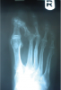
• If so, how does the metatarsal become long? It is our opinion that it is a radiographic appearance of biomechanical faults and the position of the foot in relation to the X-ray beam.
• In the case of pes cavus, does the metatarsal look short (divergent from the weightbearing surface)?
• In cases of pes planus, doesn’t the metatarsal lie more parallel to the ground, leaving an impression of a long metatarsal? In reality, the metatarsal does not become short or long after skeletal maturity.
Another common thought is that the metatarsal is “plantarflexed.” In cases in which there was no previous history of trauma or surgery, this theory is also flawed. When a fully weightbearing patient has intact deep transverse intermetatarsal ligaments along with intact interosseous musculature (lumbricales and interossei), the deep transverse intermetatarsal ligament acts like a tie-bar system. Therefore, the metatarsal cannot plantarflex.23 Based on this information, it is our opinion that second metatarsal osteotomies are not needed.
We prefer a modified Girdlestone-Taylor procedure with an intact flexor digitorum longus tendon transfer.20 We prefer this biomechanical approach to stabilization of the second metatarsophalangeal joint and researchers have well documented the results of treatment with this method.21 In cases of the “crossover hammertoe,” the extensor is involved. In these cases, we also perform a modified Hibbs procedure (localized to the second digit only), which functions as a joint sparing procedure via release of the MPJ contractures via capsulotomy and transfer of the extensor digitorum brevis tendon into the distal stump of the extensor digitorum longus tendon.20
In a study of 44 patients by Ross and Faux, the authors’ results concluded that a combined flexor-to-extensor transfer and shortening phalangeal osteotomy is an effective means of reliably stabilizing the unstable lesser MPJ.22 As we described previously, the deformity does not pertain to the bone and we have noted continued success without performing any type of digital osseous procedure.
With respect to the theory that the second metatarsal is involved, we ask the following questions:
• When does a metatarsal become “long”? Are not most of these patients adults who have reached skeletal maturity?
• If so, how does the metatarsal become long? It is our opinion that it is a radiographic appearance of biomechanical faults and the position of the foot in relation to the X-ray beam.
• In the case of pes cavus, does the metatarsal look short (divergent from the weightbearing surface)?
• In cases of pes planus, doesn’t the metatarsal lie more parallel to the ground, leaving an impression of a long metatarsal? In reality, the metatarsal does not become short or long after skeletal maturity.
Another common thought is that the metatarsal is “plantarflexed.” In cases in which there was no previous history of trauma or surgery, this theory is also flawed. When a fully weightbearing patient has intact deep transverse intermetatarsal ligaments along with intact interosseous musculature (lumbricales and interossei), the deep transverse intermetatarsal ligament acts like a tie-bar system. Therefore, the metatarsal cannot plantarflex.23 Based on this information, it is our opinion that second metatarsal osteotomies are not needed.
 If instability of the tarsal metatarsal and posterior muscle tightness exist, we will also perform a modified Lapidus with shear strain autologous calcaneal bone graft in combination with a gastrocnemius recession. The goal of the Lapidus is to restore the appropriate amount of weight under the first metatarsal and the two sesamoids. The result of performing a gastrocnemius recession in patients who present with a tight posterior muscle group is to unload the forefoot pressures (lesser metatarsals).
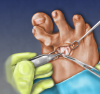
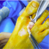
 When performing the modified Hibbs procedure for a crossover hammertoe, one can make an incision that is approximately 2 to 3 cm. Begin the incision at the second MPJ and direct it proximal and lateral. Deepen the incision in the same plane to the level of the extensor digitorum longus tendon. Lateral to the extensor digitorum longus tendon lies the smaller extensor digitorum brevis tendon. Transect the extensor digitorum longus as far as proximal in the incision site and transect the extensor digitorum brevis as far as distal in the incision site.
At this time, one can appreciate fantastic exposure to the second MPJ. The exposure of the MPJ will allow the surgeon to perform a complete release of the contracted and fibrous/deformed capsular tissue. At this time, remove all deforming forces of the MPJ. This facilitates the release of all contractures via sharp dissection and a McGlamry elevator, which allows for anatomic restoration of the MPJ. Essentially, the toe should be relaxed into a neutral “limp position” as all the deforming forces of the second MPJ pathology are gone.
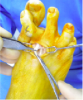
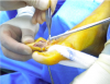
Direct your attention to the medial aspect of the second digit where you can perform a modified Girdlestone-Taylor procedure. Use a midline incision approach on the medial aspect of the toe. We recommend that the surgeon use fine double-prong skin hooks for retraction in order to avoid soft tissue compromise.
Then deepen the incision in the same plane, taking care to avoid the neurovascular bundles. Carry the incision deep to identify the flexor digitorum longus and trace it distally to its insertion of the distal phalanx. Detach the distal insertion of the flexor digitorum longus from the distal phalanx and direct it proximal to the web space.
When performing the modified Hibbs procedure for a crossover hammertoe, one can make an incision that is approximately 2 to 3 cm. Begin the incision at the second MPJ and direct it proximal and lateral. Deepen the incision in the same plane to the level of the extensor digitorum longus tendon. Lateral to the extensor digitorum longus tendon lies the smaller extensor digitorum brevis tendon. Transect the extensor digitorum longus as far as proximal in the incision site and transect the extensor digitorum brevis as far as distal in the incision site.
At this time, one can appreciate fantastic exposure to the second MPJ. The exposure of the MPJ will allow the surgeon to perform a complete release of the contracted and fibrous/deformed capsular tissue. At this time, remove all deforming forces of the MPJ. This facilitates the release of all contractures via sharp dissection and a McGlamry elevator, which allows for anatomic restoration of the MPJ. Essentially, the toe should be relaxed into a neutral “limp position” as all the deforming forces of the second MPJ pathology are gone.
Direct your attention to the medial aspect of the second digit where you can perform a modified Girdlestone-Taylor procedure. Use a midline incision approach on the medial aspect of the toe. We recommend that the surgeon use fine double-prong skin hooks for retraction in order to avoid soft tissue compromise.
Then deepen the incision in the same plane, taking care to avoid the neurovascular bundles. Carry the incision deep to identify the flexor digitorum longus and trace it distally to its insertion of the distal phalanx. Detach the distal insertion of the flexor digitorum longus from the distal phalanx and direct it proximal to the web space.
 Proceed to direct your attention to the flexor digitorum brevis tendon. Perform a tenotomy (both the medial and lateral slips) and capsulotomy at the interphalangeal joint (for a flexion contracture of the proximal interphalangeal joint). If the distal interphalangeal joint is contracted, perform a capsulotomy there as well. At this time, remove all flexion and extension contractures as well as the deforming forces from the second digit and MPJ.
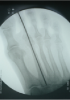
At this time, the surgeon can place the second toe into the desired anatomic position and insert a 0.062-inch Kirschner wire to stabilize and align the digit in the corrected position. Insert the K-wire from the distal tip of the distal phalanx to the base of the second metatarsal. With all the deforming forces gone and the digit in the desired anatomic position (in relation to the metatarsal), suture the flexor digitorum longus to the extensor hood under physiologic tension. It is imperative to suture this under physiologic tension as this tendon transfer will assist the lumbricales with plantarflexion and allow the toe to purchase the ground postoperatively.
Direct your attention back to the dorsum of the second MPJ and transfer the distal stump of the proximal end of the extensor digitorum brevis tendon into the proximal end of the distal stump of the extensor digitorum longus tendon via a weave graft under physiologic tension. Again, it is important to do this under physiologic tension in order to allow for the extensor digitorum brevis to dorsiflex the toe. Essentially, the modified Hibbs procedure allows the patient to maintain dorsiflexion of the toe but essentially “weakens” the dorsiflexor of the second digit.
Proceed to direct your attention to the flexor digitorum brevis tendon. Perform a tenotomy (both the medial and lateral slips) and capsulotomy at the interphalangeal joint (for a flexion contracture of the proximal interphalangeal joint). If the distal interphalangeal joint is contracted, perform a capsulotomy there as well. At this time, remove all flexion and extension contractures as well as the deforming forces from the second digit and MPJ.
At this time, the surgeon can place the second toe into the desired anatomic position and insert a 0.062-inch Kirschner wire to stabilize and align the digit in the corrected position. Insert the K-wire from the distal tip of the distal phalanx to the base of the second metatarsal. With all the deforming forces gone and the digit in the desired anatomic position (in relation to the metatarsal), suture the flexor digitorum longus to the extensor hood under physiologic tension. It is imperative to suture this under physiologic tension as this tendon transfer will assist the lumbricales with plantarflexion and allow the toe to purchase the ground postoperatively.
Direct your attention back to the dorsum of the second MPJ and transfer the distal stump of the proximal end of the extensor digitorum brevis tendon into the proximal end of the distal stump of the extensor digitorum longus tendon via a weave graft under physiologic tension. Again, it is important to do this under physiologic tension in order to allow for the extensor digitorum brevis to dorsiflex the toe. Essentially, the modified Hibbs procedure allows the patient to maintain dorsiflexion of the toe but essentially “weakens” the dorsiflexor of the second digit.
 The modified Hibbs procedure is indicated for patients who exhibit isolated extensor substitution/recruitment or global extensor substitution/recruitment to the forefoot. Dorsal subluxations/dislocations at the MPJ are frequently linked with claw toes and hammertoes, including the crossover toe variety.
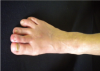
In terms of advantages with this proposed approach, the scars with the Girdlestone-Taylor procedure are located on the medial aspect of the second digit so the procedure leaves a much more cosmetically pleasing result. There is also a much more natural appearance to the digits postoperatively. Additionally, no shortening occurs and the medial and lateral collateral ligaments remain intact so no frontal plane or transverse plane complications can occur. The cubic volume of bone is not altered so instability and shortening cannot occur. Lastly, because one limits dissection to soft tissue only, the postoperative edema is minimal in relation to bony procedures.24 Of note, any resulting bursa, hyperkeratosis and/or ulceration eventually dissipate without specifically addressing them surgically. This is because the surgeon has corrected the deforming forces and relieved the abnormal pressures.
The modified Hibbs procedure is indicated for patients who exhibit isolated extensor substitution/recruitment or global extensor substitution/recruitment to the forefoot. Dorsal subluxations/dislocations at the MPJ are frequently linked with claw toes and hammertoes, including the crossover toe variety.
In terms of advantages with this proposed approach, the scars with the Girdlestone-Taylor procedure are located on the medial aspect of the second digit so the procedure leaves a much more cosmetically pleasing result. There is also a much more natural appearance to the digits postoperatively. Additionally, no shortening occurs and the medial and lateral collateral ligaments remain intact so no frontal plane or transverse plane complications can occur. The cubic volume of bone is not altered so instability and shortening cannot occur. Lastly, because one limits dissection to soft tissue only, the postoperative edema is minimal in relation to bony procedures.24 Of note, any resulting bursa, hyperkeratosis and/or ulceration eventually dissipate without specifically addressing them surgically. This is because the surgeon has corrected the deforming forces and relieved the abnormal pressures.
 Coughlin first introduced the term “second crossover toe” in 1987 to characterize a multi-plane deformity at the metatarsophalangeal joint (MPJ).1 Second MPJ instability is all too often a point of consternation for the foot and ankle surgeon.
Crossover toe deformity commonly results from instability of the second MPJ in conjunction with further weakness resulting from hallux abductovalgus deformity and often gastrocnemius equinus. Instability occurs in the sagittal plane but in terms of the crossover toe, this is also combined with a subluxation and/or dislocation in a dorsal and medial direction.2 Once the second MPJ is overloaded in a chronic state, plantar plate rupture and collateral ligament damage can also occur. Further weakened by hallux abductovalgus deformity, instability of the second MPJ can ultimately lead to crossover toe deformity.
Proper treatment of this condition has long been a topic of debate. Regardless, it is paramount to address the underlying biomechanical factors. One should also be cognizant that this is not a result of bony pathology and surgeons can achieve suitable correction through soft tissue balancing procedures.
Coughlin first introduced the term “second crossover toe” in 1987 to characterize a multi-plane deformity at the metatarsophalangeal joint (MPJ).1 Second MPJ instability is all too often a point of consternation for the foot and ankle surgeon.
Crossover toe deformity commonly results from instability of the second MPJ in conjunction with further weakness resulting from hallux abductovalgus deformity and often gastrocnemius equinus. Instability occurs in the sagittal plane but in terms of the crossover toe, this is also combined with a subluxation and/or dislocation in a dorsal and medial direction.2 Once the second MPJ is overloaded in a chronic state, plantar plate rupture and collateral ligament damage can also occur. Further weakened by hallux abductovalgus deformity, instability of the second MPJ can ultimately lead to crossover toe deformity.
Proper treatment of this condition has long been a topic of debate. Regardless, it is paramount to address the underlying biomechanical factors. One should also be cognizant that this is not a result of bony pathology and surgeons can achieve suitable correction through soft tissue balancing procedures.
Understanding The Possible Etiological Factors Of Crossover Hammertoe
Common surgical procedures for the correction of the second MPJ abnormalities include single tenodesis techniques, such as flexor-to-extensor tendon transfer, a variety of metatarsal osteotomies, digital arthroplasty or arthrodesis, second MPJ arthrodesis, direct plantar plate repair, and even amputation.3-9 When it comes to second MPJ instability, the literature notes several etiologic factors including seronegative and seropositive rheumatoid arthritis as well as hypermobility (e.g. Ehlers-Danlos syndrome).1 Lesser MPJ instability can be associated with hallux valgus, hallux rigidus, interdigital neuromas and hammertoe deformities.10 Regardless of pathology, authors have described the plantar plate as the most significant factor in stabilizing the MPJ.7 In the early stages of the deformity, the diagnosis can be challenging with a second intermetatarsal space neuroma and synovial cyst being included in the forefront of the differential diagnoses.2,5 Despite the name “crossover hammertoe,” the misalignment does not always progress to crossing over or under the adjacent digit until later stages, and can also affect other lesser digits besides the second digit.10 However, the second MPJ is the most common chronically dislocated joint in the foot so we will focus on that.7 There are several etiological theories as to the underlying etiology of the subluxed/dislocated second MPJ.7 Acute trauma or, more commonly, chronic microtrauma to the MPJ can cause disruption and deterioration of the plantar plate, joint capsule and collateral ligaments, leading to instability.11 Yu and colleagues originally coined early stage MPJ instability as predislocation syndrome.12 Crossover toes represent an end-stage deformity with the abnormality being previously noted in the plantar plate as well as in the collateral ligaments of the lesser MPJ complex.8
When examining the patient with a crossover toe, one should scrutinize the presence of a hallux valgus deformity, digital contracture, plantarflexed second metatarsal, second MPJ instability, neuritic symptoms and gastrocnemius contracture.10 The gastrocnemius muscle is the predominant deforming force in patients with structural breakdown and chronic pathological changes related to the foot and ankle.13 Therefore, we stress the importance of addressing equinus if any surgical correction is going to be successful.
There are several etiological theories as to the underlying etiology of the subluxed/dislocated second MPJ.7 Acute trauma or, more commonly, chronic microtrauma to the MPJ can cause disruption and deterioration of the plantar plate, joint capsule and collateral ligaments, leading to instability.11 Yu and colleagues originally coined early stage MPJ instability as predislocation syndrome.12 Crossover toes represent an end-stage deformity with the abnormality being previously noted in the plantar plate as well as in the collateral ligaments of the lesser MPJ complex.8
When examining the patient with a crossover toe, one should scrutinize the presence of a hallux valgus deformity, digital contracture, plantarflexed second metatarsal, second MPJ instability, neuritic symptoms and gastrocnemius contracture.10 The gastrocnemius muscle is the predominant deforming force in patients with structural breakdown and chronic pathological changes related to the foot and ankle.13 Therefore, we stress the importance of addressing equinus if any surgical correction is going to be successful.
A Closer Look At Tendon Transfers And Juxtaposed Procedures
A crossover toe characteristically presents in a varus position at the MPJ, resulting in a hammertoe deformity. This intrinsic muscular instability is a result of a mechanical disadvantage that typically occurs at the second MPJ and is coupled with a hallux valgus deformity.14 The second MPJ is frequently subject to increased weightbearing loads, which produce progressive attenuation of the plantar plate insertion into the proximal phalanx. In patients with a combined hallux valgus deformity, this further increases the load placed on the second metatarsal head.6 It is helpful to remember that studies show 60 percent of normal weightbearing forces pass through the first ray from heel strike to toe-off.15 Once the first ray fails to support this necessary load, the medial column collapses. This also leads to a lateral load shift to the lesser metatarsals, continued biomechanical abnormalities and continued deforming forces, perpetuating an aggressive cycle. Likewise, Morton proposed that the unstable first ray is an inherent cause of second metatarsal pathology.16 Greisberg and coworkers also reported similar findings as patients with second MPJ synovitis and metatarsalgia had greater first ray mobility. In clinical situations, the severe bunion is associated with lesser metatarsal overload (metatarsalgia) through an inefficient medial column (first ray).17 Stabilizing and realigning the first ray through a Lapidus procedure provides a stable construct to the medial column and also improves the efficiency of the peroneus longus.18 Following the Lapidus arthrodesis, the first ray is better able to absorb the ground reactive forces during weightbearing. The procedure is predictable in stabilizing the first ray in three planes with emphasis on the sagittal and frontal planes along with improved intermetatarsal angles.19
Crossover toe deformity treatment goals are to reduce the deformity, provide pain relief, improve function, reduce morbidity and prevent the progression of the existing deformity. The surgical approach to digital crossover deformity varies and is based on patient suitability and the surgeon’s preference. One may modify the surgical approach to address the presentation of contracted digits with or without additional MPJ pathology, hallux abductovalgus and first ray stability.20
The digital joint-destructive procedures include arthroplasty and arthrodesis as well as digital implants. Joint-sparing hammertoe correction is based on soft-tissue rebalancing and may entail open, percutaneous or closed procedures that aim at reducing capsular and tendon contractures, and deforming soft tissue forces by balancing the tendons.20
Likewise, Morton proposed that the unstable first ray is an inherent cause of second metatarsal pathology.16 Greisberg and coworkers also reported similar findings as patients with second MPJ synovitis and metatarsalgia had greater first ray mobility. In clinical situations, the severe bunion is associated with lesser metatarsal overload (metatarsalgia) through an inefficient medial column (first ray).17 Stabilizing and realigning the first ray through a Lapidus procedure provides a stable construct to the medial column and also improves the efficiency of the peroneus longus.18 Following the Lapidus arthrodesis, the first ray is better able to absorb the ground reactive forces during weightbearing. The procedure is predictable in stabilizing the first ray in three planes with emphasis on the sagittal and frontal planes along with improved intermetatarsal angles.19
Crossover toe deformity treatment goals are to reduce the deformity, provide pain relief, improve function, reduce morbidity and prevent the progression of the existing deformity. The surgical approach to digital crossover deformity varies and is based on patient suitability and the surgeon’s preference. One may modify the surgical approach to address the presentation of contracted digits with or without additional MPJ pathology, hallux abductovalgus and first ray stability.20
The digital joint-destructive procedures include arthroplasty and arthrodesis as well as digital implants. Joint-sparing hammertoe correction is based on soft-tissue rebalancing and may entail open, percutaneous or closed procedures that aim at reducing capsular and tendon contractures, and deforming soft tissue forces by balancing the tendons.20
 We prefer a modified Girdlestone-Taylor procedure with an intact flexor digitorum longus tendon transfer.20 We prefer this biomechanical approach to stabilization of the second metatarsophalangeal joint and researchers have well documented the results of treatment with this method.21 In cases of the “crossover hammertoe,” the extensor is involved. In these cases, we also perform a modified Hibbs procedure (localized to the second digit only), which functions as a joint sparing procedure via release of the MPJ contractures via capsulotomy and transfer of the extensor digitorum brevis tendon into the distal stump of the extensor digitorum longus tendon.20
In a study of 44 patients by Ross and Faux, the authors’ results concluded that a combined flexor-to-extensor transfer and shortening phalangeal osteotomy is an effective means of reliably stabilizing the unstable lesser MPJ.22 As we described previously, the deformity does not pertain to the bone and we have noted continued success without performing any type of digital osseous procedure.
With respect to the theory that the second metatarsal is involved, we ask the following questions:
• When does a metatarsal become “long”? Are not most of these patients adults who have reached skeletal maturity?
• If so, how does the metatarsal become long? It is our opinion that it is a radiographic appearance of biomechanical faults and the position of the foot in relation to the X-ray beam.
• In the case of pes cavus, does the metatarsal look short (divergent from the weightbearing surface)?
• In cases of pes planus, doesn’t the metatarsal lie more parallel to the ground, leaving an impression of a long metatarsal? In reality, the metatarsal does not become short or long after skeletal maturity.
Another common thought is that the metatarsal is “plantarflexed.” In cases in which there was no previous history of trauma or surgery, this theory is also flawed. When a fully weightbearing patient has intact deep transverse intermetatarsal ligaments along with intact interosseous musculature (lumbricales and interossei), the deep transverse intermetatarsal ligament acts like a tie-bar system. Therefore, the metatarsal cannot plantarflex.23 Based on this information, it is our opinion that second metatarsal osteotomies are not needed.
We prefer a modified Girdlestone-Taylor procedure with an intact flexor digitorum longus tendon transfer.20 We prefer this biomechanical approach to stabilization of the second metatarsophalangeal joint and researchers have well documented the results of treatment with this method.21 In cases of the “crossover hammertoe,” the extensor is involved. In these cases, we also perform a modified Hibbs procedure (localized to the second digit only), which functions as a joint sparing procedure via release of the MPJ contractures via capsulotomy and transfer of the extensor digitorum brevis tendon into the distal stump of the extensor digitorum longus tendon.20
In a study of 44 patients by Ross and Faux, the authors’ results concluded that a combined flexor-to-extensor transfer and shortening phalangeal osteotomy is an effective means of reliably stabilizing the unstable lesser MPJ.22 As we described previously, the deformity does not pertain to the bone and we have noted continued success without performing any type of digital osseous procedure.
With respect to the theory that the second metatarsal is involved, we ask the following questions:
• When does a metatarsal become “long”? Are not most of these patients adults who have reached skeletal maturity?
• If so, how does the metatarsal become long? It is our opinion that it is a radiographic appearance of biomechanical faults and the position of the foot in relation to the X-ray beam.
• In the case of pes cavus, does the metatarsal look short (divergent from the weightbearing surface)?
• In cases of pes planus, doesn’t the metatarsal lie more parallel to the ground, leaving an impression of a long metatarsal? In reality, the metatarsal does not become short or long after skeletal maturity.
Another common thought is that the metatarsal is “plantarflexed.” In cases in which there was no previous history of trauma or surgery, this theory is also flawed. When a fully weightbearing patient has intact deep transverse intermetatarsal ligaments along with intact interosseous musculature (lumbricales and interossei), the deep transverse intermetatarsal ligament acts like a tie-bar system. Therefore, the metatarsal cannot plantarflex.23 Based on this information, it is our opinion that second metatarsal osteotomies are not needed.
 If instability of the tarsal metatarsal and posterior muscle tightness exist, we will also perform a modified Lapidus with shear strain autologous calcaneal bone graft in combination with a gastrocnemius recession. The goal of the Lapidus is to restore the appropriate amount of weight under the first metatarsal and the two sesamoids. The result of performing a gastrocnemius recession in patients who present with a tight posterior muscle group is to unload the forefoot pressures (lesser metatarsals).
Pertinent Pearls Of The Authors’ Surgical Technique
Most crossover digit deformities are associated with an abnormal pull and biomechanics of the short and long flexors and extensors that have caused the toe to deform. The surgery must balance the flexors and extensors in order to prevent recurrence or continued progression of the deformity. As a matter of fact, once one removes the deforming force, a recurrence cannot happen. Opting for soft tissue techniques involving the short and long flexors and extensors may be more beneficial, especially in patients with significant compromise of the plantar plate. We do not think that plantar plate repairs or arthroplasties/arthrodesis are the indicated treatment choice to address this problem. Lastly, if the attempted soft tissue procedures would not succeed, the surgeon could always revert back to a bony procedure. If the surgeon appropriately addresses the multiplanar crossover deformity, he or she should be able to correct all planes of deformity. A modified Girdlestone-Taylor procedure can transfer the flexor digitorum longus tendon to the extensor hood. A modified Hibbs procedure will release the extensor medial and varus pull of the pathologic position. The modified Hibbs procedure can decrease the extensor tendon retrograde buckling of the second digit on the second metatarsal. Furthermore, this release will allow the surgeon to mobilize the crossover toe out of the varus and medially directed position while providing excellent exposure to the metatarsophalangeal joint. When performing the modified Hibbs procedure for a crossover hammertoe, one can make an incision that is approximately 2 to 3 cm. Begin the incision at the second MPJ and direct it proximal and lateral. Deepen the incision in the same plane to the level of the extensor digitorum longus tendon. Lateral to the extensor digitorum longus tendon lies the smaller extensor digitorum brevis tendon. Transect the extensor digitorum longus as far as proximal in the incision site and transect the extensor digitorum brevis as far as distal in the incision site.
At this time, one can appreciate fantastic exposure to the second MPJ. The exposure of the MPJ will allow the surgeon to perform a complete release of the contracted and fibrous/deformed capsular tissue. At this time, remove all deforming forces of the MPJ. This facilitates the release of all contractures via sharp dissection and a McGlamry elevator, which allows for anatomic restoration of the MPJ. Essentially, the toe should be relaxed into a neutral “limp position” as all the deforming forces of the second MPJ pathology are gone.
Direct your attention to the medial aspect of the second digit where you can perform a modified Girdlestone-Taylor procedure. Use a midline incision approach on the medial aspect of the toe. We recommend that the surgeon use fine double-prong skin hooks for retraction in order to avoid soft tissue compromise.
Then deepen the incision in the same plane, taking care to avoid the neurovascular bundles. Carry the incision deep to identify the flexor digitorum longus and trace it distally to its insertion of the distal phalanx. Detach the distal insertion of the flexor digitorum longus from the distal phalanx and direct it proximal to the web space.
When performing the modified Hibbs procedure for a crossover hammertoe, one can make an incision that is approximately 2 to 3 cm. Begin the incision at the second MPJ and direct it proximal and lateral. Deepen the incision in the same plane to the level of the extensor digitorum longus tendon. Lateral to the extensor digitorum longus tendon lies the smaller extensor digitorum brevis tendon. Transect the extensor digitorum longus as far as proximal in the incision site and transect the extensor digitorum brevis as far as distal in the incision site.
At this time, one can appreciate fantastic exposure to the second MPJ. The exposure of the MPJ will allow the surgeon to perform a complete release of the contracted and fibrous/deformed capsular tissue. At this time, remove all deforming forces of the MPJ. This facilitates the release of all contractures via sharp dissection and a McGlamry elevator, which allows for anatomic restoration of the MPJ. Essentially, the toe should be relaxed into a neutral “limp position” as all the deforming forces of the second MPJ pathology are gone.
Direct your attention to the medial aspect of the second digit where you can perform a modified Girdlestone-Taylor procedure. Use a midline incision approach on the medial aspect of the toe. We recommend that the surgeon use fine double-prong skin hooks for retraction in order to avoid soft tissue compromise.
Then deepen the incision in the same plane, taking care to avoid the neurovascular bundles. Carry the incision deep to identify the flexor digitorum longus and trace it distally to its insertion of the distal phalanx. Detach the distal insertion of the flexor digitorum longus from the distal phalanx and direct it proximal to the web space.
 Proceed to direct your attention to the flexor digitorum brevis tendon. Perform a tenotomy (both the medial and lateral slips) and capsulotomy at the interphalangeal joint (for a flexion contracture of the proximal interphalangeal joint). If the distal interphalangeal joint is contracted, perform a capsulotomy there as well. At this time, remove all flexion and extension contractures as well as the deforming forces from the second digit and MPJ.
At this time, the surgeon can place the second toe into the desired anatomic position and insert a 0.062-inch Kirschner wire to stabilize and align the digit in the corrected position. Insert the K-wire from the distal tip of the distal phalanx to the base of the second metatarsal. With all the deforming forces gone and the digit in the desired anatomic position (in relation to the metatarsal), suture the flexor digitorum longus to the extensor hood under physiologic tension. It is imperative to suture this under physiologic tension as this tendon transfer will assist the lumbricales with plantarflexion and allow the toe to purchase the ground postoperatively.
Direct your attention back to the dorsum of the second MPJ and transfer the distal stump of the proximal end of the extensor digitorum brevis tendon into the proximal end of the distal stump of the extensor digitorum longus tendon via a weave graft under physiologic tension. Again, it is important to do this under physiologic tension in order to allow for the extensor digitorum brevis to dorsiflex the toe. Essentially, the modified Hibbs procedure allows the patient to maintain dorsiflexion of the toe but essentially “weakens” the dorsiflexor of the second digit.
Proceed to direct your attention to the flexor digitorum brevis tendon. Perform a tenotomy (both the medial and lateral slips) and capsulotomy at the interphalangeal joint (for a flexion contracture of the proximal interphalangeal joint). If the distal interphalangeal joint is contracted, perform a capsulotomy there as well. At this time, remove all flexion and extension contractures as well as the deforming forces from the second digit and MPJ.
At this time, the surgeon can place the second toe into the desired anatomic position and insert a 0.062-inch Kirschner wire to stabilize and align the digit in the corrected position. Insert the K-wire from the distal tip of the distal phalanx to the base of the second metatarsal. With all the deforming forces gone and the digit in the desired anatomic position (in relation to the metatarsal), suture the flexor digitorum longus to the extensor hood under physiologic tension. It is imperative to suture this under physiologic tension as this tendon transfer will assist the lumbricales with plantarflexion and allow the toe to purchase the ground postoperatively.
Direct your attention back to the dorsum of the second MPJ and transfer the distal stump of the proximal end of the extensor digitorum brevis tendon into the proximal end of the distal stump of the extensor digitorum longus tendon via a weave graft under physiologic tension. Again, it is important to do this under physiologic tension in order to allow for the extensor digitorum brevis to dorsiflex the toe. Essentially, the modified Hibbs procedure allows the patient to maintain dorsiflexion of the toe but essentially “weakens” the dorsiflexor of the second digit.
 The modified Hibbs procedure is indicated for patients who exhibit isolated extensor substitution/recruitment or global extensor substitution/recruitment to the forefoot. Dorsal subluxations/dislocations at the MPJ are frequently linked with claw toes and hammertoes, including the crossover toe variety.
In terms of advantages with this proposed approach, the scars with the Girdlestone-Taylor procedure are located on the medial aspect of the second digit so the procedure leaves a much more cosmetically pleasing result. There is also a much more natural appearance to the digits postoperatively. Additionally, no shortening occurs and the medial and lateral collateral ligaments remain intact so no frontal plane or transverse plane complications can occur. The cubic volume of bone is not altered so instability and shortening cannot occur. Lastly, because one limits dissection to soft tissue only, the postoperative edema is minimal in relation to bony procedures.24 Of note, any resulting bursa, hyperkeratosis and/or ulceration eventually dissipate without specifically addressing them surgically. This is because the surgeon has corrected the deforming forces and relieved the abnormal pressures.
The modified Hibbs procedure is indicated for patients who exhibit isolated extensor substitution/recruitment or global extensor substitution/recruitment to the forefoot. Dorsal subluxations/dislocations at the MPJ are frequently linked with claw toes and hammertoes, including the crossover toe variety.
In terms of advantages with this proposed approach, the scars with the Girdlestone-Taylor procedure are located on the medial aspect of the second digit so the procedure leaves a much more cosmetically pleasing result. There is also a much more natural appearance to the digits postoperatively. Additionally, no shortening occurs and the medial and lateral collateral ligaments remain intact so no frontal plane or transverse plane complications can occur. The cubic volume of bone is not altered so instability and shortening cannot occur. Lastly, because one limits dissection to soft tissue only, the postoperative edema is minimal in relation to bony procedures.24 Of note, any resulting bursa, hyperkeratosis and/or ulceration eventually dissipate without specifically addressing them surgically. This is because the surgeon has corrected the deforming forces and relieved the abnormal pressures.

