A Novel Technique to Anesthetize the Skin Without Obscuring a Deeply Palpable Lesion
In a typically idealized circumstance of lesion removal, the dermatologic surgeon initially positions the patient in the standard surgical fashion, palpates the lesion to the best extent possible, identifies the circumscribed borders of the lesion, and then proceeds to mark the proposed incision site of the skin overlying the palpable lesion. This is followed by a wheal infiltration of the incision site to anesthetize the surgical soft tissue. However, a subset of dermatologic procedures involves the surgical extrication of subcutaneous nodules that are barely palpable under the skin. Examples include lesions that are mobile and poorly circumscribed, as seen with foreign body material, lymph nodes, or subfascial tumors. In these cases, the infiltration of local anesthetic into the skin and soft tissue prior to dermatologic surgery may obfuscate the borders of the lesion, making it more difficult to identify the targeted pathology.
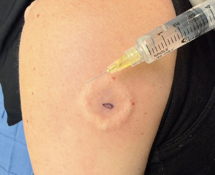
We propose a novel methodology that can be used to anesthetize the surgical soft tissue footprint when the dermatologic surgeon identifies a subcutaneous lesion that is inherently difficult to palpate and localize prior to excision. This method involves a ring block infiltration of local anesthetic along the perimeter of the identified lesion, leaving the overlying skin above the lesion unaffected.
Ring block infiltration involves dermal injection of local anesthetic in a circumferential manner along a measured radius from the central point overlying the subcutaneous lesion (Figure). Presumably, the entire footprint contained within the circular wheal will be anesthetized; this is, of course, considering that the injection site is absent of artifact swelling induced by non-strategic injection of anesthetic.
The specific limitation of the ring block infiltration technique is that although it can effectively anesthetize the entire surgical field, the vasoconstrictive effects of the epinephrine-infused anesthetic may be limited at the central incision site.
The ring block infiltration technique offers particular utility in scenarios where precise lesion localization is paramount to surgical success. Mobile subcutaneous lymph nodes, embedded foreign bodies, and deeply seated lipomas represent clinical situations where maintaining tactile feedback throughout the procedure can significantly improve excision accuracy and reduce the risk of incomplete removal. By preserving the natural tissue architecture overlying the lesion, the surgeon maintains the ability to reassess lesion position and dimensions following anesthesia administration. Implementation of this technique requires careful consideration of the anesthetic volume and radius of infiltration, with local anesthetic injected at multiple equidistant points along a circle surrounding the lesion’s estimated center. The dermal wheals should be allowed to coalesce for several minutes before initiating the procedure, ensuring adequate anesthetic diffusion to the central surgical field.
While traditional field block or tumescent anesthesia techniques may be preferred for certain procedures involving superficial lesions with well-defined borders or extensive undermining, the ring block method offers distinct advantages in clinical scenarios where preserving palpable lesion boundaries is beneficial. Surgeons should consider this approach when dealing with poorly circumscribed or mobile subcutaneous pathology, and be prepared to supplement with additional anesthetic if needed during the procedure.
Kushagra Tewari is a medical student at the UCLA School of Medicine in Los Angeles, CA. Levon Karamanoukian is a student at Brentwood School in Los Angeles, CA, and a research intern for Dr Rtail. Dr Rtail is a facial plastic surgeon at Saint Joseph Medical Center in Beirut, Lebanon.
Disclosure: The authors report no relevant financial relationships.