


Plantar-to-Plantar Punch Skin Grafts for a Wound Caused by Crush Injury
© 2026 HMP Global. All Rights Reserved.
Any views and opinions expressed are those of the author(s) and/or participants and do not necessarily reflect the views, policy, or position of Wounds or HMP Global, their employees, and affiliates.
Abstract
Background. Skin grafting of the sole of the foot poses a special challenge because it is subjected to friction and shear. Various kinds of grafts have been used throughout history for this purpose based on the features of the donor site and graft thickness. Given these unique characteristics, as well as the need to withstand pressure bearing and friction, the best grafts should be obtained from identical skin. Case Report. A patient with a crush wound of the plantar and dorsal region of the left foot underwent combined treatment with plantar-to-plantar punch grafting and split-thickness skin grafting of the dorsal region to obtain full closure with appropriate region-specific skin. One week later, 85% of the grafts had taken and the donor site was healing. The patient refused a second grafting procedure and decided to wait for the initial grafts to continue growing. After 3 months, complete healing of both the dorsal and the plantar aspects of the foot was achieved. Conclusion. Skin obtained from other sites lacks, and will not develop, the same phenotype as plantar skin, which may result in complications such as hair growth, re-ulceration, hyperkeratosis, marginal scarring, pain, hyperpigmentation, or recurrent breakdown of a preexisting graft. This case demonstrates success with grafting skin of the same glabrous phenotype.
It was Jacques-Louis Reverdin who in 1869 first utilized punch grafting as a means of closing skin defects.1 Autografts have evolved since then and remain a reconstructive option; they are sometimes preferred for complex wounds. In reconstructive surgery, “replacing like with like” should always be the guiding principle.2
The glabrous skin of the sole of the foot has unique characteristics that enable it to perform its functions. These include a thick epidermal layer with a solid stratum corneum, a thick stratum granulosum, a well-defined stratum lucidum to resist shearing forces and pressure, a compact dermis with reduced elasticity, absence of melanocytes and hair follicles, abundant sweat glands that provide moisture, and numerous pacinian corpuscles for optimal sensibility.3
Given these special features, grafting skin of a different phenotype (ie, nonglabrous skin) can result in multiple complications, such as re-ulceration due to differences in thickness and structural resilience under daily friction and pressure.3 Use of more elastic skin can lead to contraction, causing functional limitations, as well as color mismatch or unwanted hair growth, as has been described in the treatment of palm injuries with grafts from skin of a different phenotype.4-6 An advantage of using glabrous skin includes the abundance of sweat glands, which support multicentric epithelial budding and provide the humidity necessary for faster healing of both donor and recipient sites compared with conventional nonglabrous skin grafts.3
The punch graft technique provides multiple full-thickness skin grafts using relatively small areas of the sole; these grafts can be used to repair defects on either the ipsilateral or the contralateral foot. This technique is easy to learn, is cost-effective, and can be performed on an outpatient basis without the need for an operating room.7 However, its use may be limited by the availability of healthy skin in cases of large foot defects. Whenever possible, skin from non–weight-bearing areas of the foot is preferred.8
The authors present the case of a patient who sustained a crush injury to the foot with degloving caused by ischemic necrosis, and who was treated with a combined approach comprising plantar-
to-plantar punch grafts to reconstruct the sole and split-thickness skin grafts for coverage of the dorsum.
Case Report
A 54-year-old male with no relevant medical history presented on September 14, 2020, with ulceration and necrosis of the left foot secondary to a crush injury caused by a 30-ton metal sheet bender 2 weeks earlier (Figure 1). Below-knee amputation was the only treatment option previously offered, and he sought a second opinion.
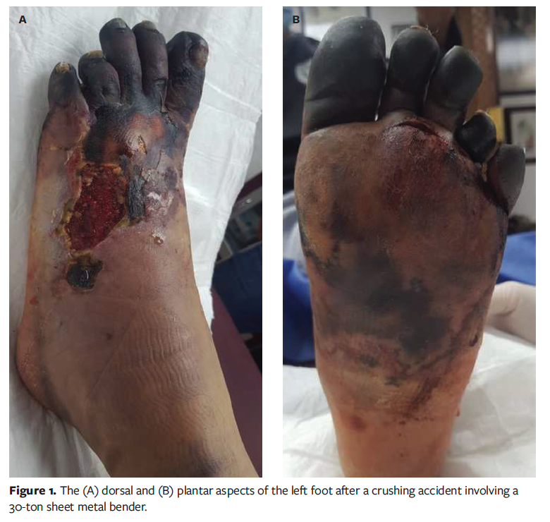
Physical examination revealed necrosis of the toes with detachment of the distal phalanges. Debridement and drainage of a large hematoma on the plantar aspect of the foot were performed, resulting in a large defect extending from the dorsum to the plantar midfoot. No signs of infection were observed. The wounds were initially managed with calcium alginate containing silver (Algisite Ag; Smith & Nephew), and 3 sessions of dry wound therapy were prescribed for the remaining necrotic areas. During follow-up visits, regular debridement and wound care promoted granulation tissue formation.
Two months after initial debridement and drainage, partial salvage of the first toe and several phalanges was achieved; however, a large ulcer persisted, affecting both the dorsal and the plantar aspect of the foot (Figure 2A, 2B).
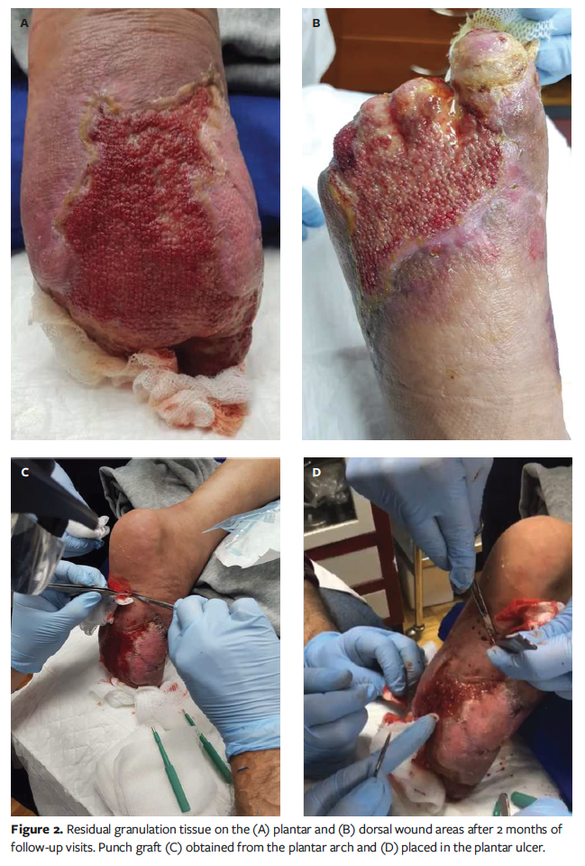
Grafting was subsequently performed under local anesthesia. For the plantar ulcer, 32 full-thickness, 4-mm punch grafts were harvested from the remaining skin over the arch and placed over the defect (Figure 2C, 2D). Using a safety blade (Personna DermaBlade; AccuThrive), split-thickness skin grafts were harvested from the medial thighs to cover the dorsal defect.
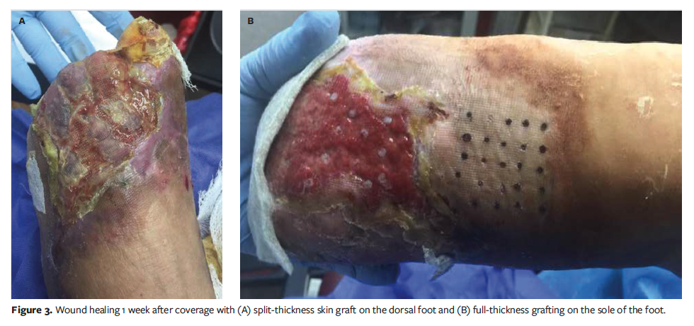
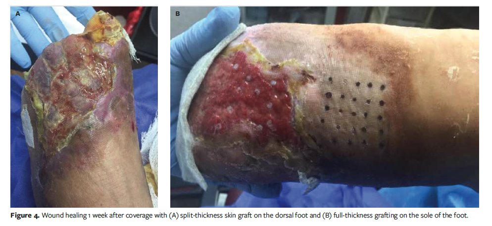
The grafts were secured using nonadhesive gauze (Curity non-adhering dressing; Cardinal Health) and cotton gauze, and the donor sites were covered with hydrocolloid dressings (Duoderm; Convatec). Both areas were bandaged with compression. One week later, 85% of the grafts had taken, and the donor sites demonstrated healing (Figure 3). The patient declined a second grafting procedure, opting instead to allow the existing grafts to continue maturing.
At 3 months, complete healing of both the dorsal and the plantar aspects of the foot was achieved (Figure 4). The patient subsequently resumed normal activities and was referred for rehabilitation.
Informed consent was received from the patient for the publication of this case report and associated images.
Discussion
Proper donor site selection should promote rapid recovery and minimal scarring, always emphasizing the principle of grafting “like with like.”2 Skin of a different phenotype will not acquire the unique characteristics of the recipient site; this is a common misconception.
Both in vitro and in vivo studies have demonstrated that site-specific differentiation of postnatal keratinocytes is an intrinsic property that is expressed and maintained after grafting. In a comparison of plantar and nonplantar cultured keratinocyte grafts in pediatric burn patients, grafts originating from plantar skin showed the typical thick stratum corneum, thick stratum granulosum, and presence of a stratum lucidum within 2 weeks of grafting.9 Notably, the formation of rete ridges was observed after 2 months in plantar grafts, compared with 4 months to 6 months in nonplantar skin grafts. In vitro, plantar keratinocytes initially expressed very low keratin, but its synthesis increased markedly after 7 days postgrafting and was maintained thereafter.9
Using nonglabrous donor sites for areas like the sole can result in complications that impair rehabilitation and ambulation, with frequent re-ulceration of the grafted areas. Additional complications include hyperpigmentation, graft contracture, hair growth, ulceration, folliculitis, hyperkeratosis, marginal scarring, and recurrent breakdown of a preexisting graft.3,5,10 In contrast, using plantar skin preserves resistance to regular friction and weight-bearing forces, thereby avoiding these outcomes. In a study comparing scars derived from glabrous versus nonglabrous skin, assessments using colorimetry, pigmentation scores, and the Vancouver Scar Scale (VSS) showed superior results with glabrous skin grafts.11 Another study demonstrated a notable improvement on the VSS, with average scores improving from 9.53 to 2.53, reflecting better outcomes in both color and texture.4
Punch grafting represents an excellent alternative for complex wounds of various etiologies due to its efficacy, simplicity, low cost, and rapid recovery.7 The non–weight-bearing area of the foot is always preferred as the donor site to minimize morbidity; however, the submalleolar region may also be used if needed.6 Whenever possible, grafting from the ipsilateral foot is recommended to preserve the contralateral foot for normal ambulation.7 In the present case, this technique enabled the use of a small amount of donor skin, achieving rapid recovery with excellent cosmetic results. These outcomes are consistent with previous reports, in which donor sites typically healed within 2 weeks.2,12
Whereas the epidermis is relatively simple tissue capable of regenerating fully after injury, the dermis is more complex and, in mammals, heals by forming scar tissue rather than regenerating ad integrum.13 In full-thickness wounds on the sole of the foot, complete reepithelialization rarely occurs before 4 weeks and can take up to 12 weeks to 16 weeks in complicated cases, compromising function and aesthetics by obtaining tissue without the characteristics of glabrous skin.14,15
In this patient’s case of a large ulcer with loss of both dermal and epidermal components, the use of phenotype-specific grafts for the dorsum and sole of the foot promoted superior healing with fewer complications than had grafts of a different phenotype been used.
Limitations
This case report describes the successful use of plantar punch grafts for a single patient and therefore may not be generalizable to all crush injuries or plantar wounds. Larger defects may require additional procedures or alternative grafting techniques if sufficient donor skin is unavailable. Long-term follow-up beyond 3 months was not included, and thus, durability over extended periods remains to be evaluated. Furthermore, functional outcomes such as plantar sensation and gait were not formally assessed using validated scales.
Conclusion
When reconstructing plantar defects, using skin of the same glabrous phenotype is essential to restore function, resist mechanical stress, and reduce complications such as graft breakdown, ulceration, or contracture. Punch grafting from non–weight-bearing areas of the foot offers a simple, effective, and cosmetically favorable technique, allowing for rapid healing of both donor and recipient sites. In this case, the use of plantar punch grafts combined with split-thickness skin grafts successfully restored a severely injured foot, demonstrating that phenotype-specific grafting can optimize both functional and aesthetic outcomes. This approach represents a valuable, low-cost alternative for complex foot wounds, especially when preserving ambulation and minimizing morbidity are primary goals.
Author and Public Information
Authors: Francisco Javier Ramírez-Fernández, MD1; Isidro Velázquez-Jacobo, MD2; Adriana Lozano-Platonoff, MD3; Monica Navarrete-Martínez, MD4; and José Contreras-Ruiz, MD4
Affiliations: 1Plastic, Aesthetic, and Reconstructive Surgery, “Dr. Bernardo J. Gastélum” General Hospital of Culiacán, Culiacán, Mexico; 2Mexico Wound and Ostomy Care Center, Mexico City, Mexico; 3Division of Dermatology, “Dr. Manuel Gea González” General Hospital, Mexico City, Mexico; 4Polanco Dermatology Center, Los Cabos, Baja California Sur, Mexico
Disclosures: The authors disclose no financial or other conflicts of interest.
Ethical Approval: Informed consent was received from the patient for the publication of this case report and associated images.
Correspondence: José Contreras-Ruiz, MD; Polanco Dermatology Center, Los Cabos, 13 Malecón San José, San José del Cabo, Los Cabos, Baja California Sur, Mexico, 23405; dermayheridas@gmail.com
Manuscript Accepted: October 31, 2025
References
1. Fariña-Pérez LA. Jaques-Louis Reverdin (1842-1921): El cirujano y la aguja. Arch Esp Urol. 2010;63(4):269-274.
2. Simman R. Medial plantar arch pinch grafts are an effective technique to resurface palmar and plantar wounds. Ann Plast Surg. 2004;53(3):256-260. doi:10.1097/01.sap.0000116247.68396.35
3. Robotti EB, Edstrom LE. Split-thickness plantar skin grafts for coverage in the hand and digits. J Hand Surg Am. 1991;16(1):143-146. doi:10.1016/S0363-5023(10)80032-2
4. Moon SH, Lee SY, Jung SN, et al. Use of split thickness plantar skin grafts in the treatment of hyperpigmented skin-grafted fingers and palms in previously burned patients. Burns. 2011;37(4):714-720. doi:10.1016/j.burns.2011.01.010
5. Teles G, Bastos V, Mello G. Correction of hyperchromic palmar graft with split-thickness instep plantar graft: case report. J Burn Care Res. 2008;29(2):403-405. doi:10.1097/BCR.0b013e3181667963
6. Zoltie N, Verlende P, Logan A. Full thickness grafts taken from the plantar instep for syndactyly release. J Hand Surg Br. 1989;14(2):201-203. doi:10.1016/0266-7681_89_90126-5
7. Garlatti MLB, Kowalczuk MVR, Anahi LB, et al. Injertos por punch en el tratamiento de úlceras de difícil manejo. Informe de dos casos. Dermatología Cosmética, Médica y Quirúrgica. 2018;16(1):76-82.
8. Wang CY, Chang CK, Chou CY, et al. Successful treatment of plantar hyperkeratosis in the form of recurrent corns with split-thickness sole skin graft. Ann Plast Surg. 2018;80(2S Suppl 1):S55-S58. doi:10.1097/SAP.0000000000001304
9. Compton CC, Nadire KB, Regauer S, et al. Cultured human sole-derived keratinocyte grafts re-express site-specific differentiation after transplantation. Differentiation. 1998;64(1):45-53. doi:10.1046/j.1432-0436.1998.6410045.x
10. Moon SH, Jung SN. Response to: an approach to ‘use of split thickness plantar skin grafts in the treatment of hyperpigmented skin-grafted fingers and palms in previously burned patients’. Burns. 2013;39(8):1651. doi:10.1016/j.burns.2013.07.013
11. Elrod J, Moellmeier D, Schiestl C, Mohr C, Neuhaus K. Comparative analysis of functional and aesthetic outcomes of retroauricular full thickness versus plantar glabrous split thickness skin grafts in pediatric palmar hand burns. Burns. 2020;46(3):639-646. doi:10.1016/j.burns.2019.09.004
12. Liu HH, Chang CK, Huang CH, et al. Use of split-thickness plantar skin grafts in the management of leg and foot skin defects. Int Wound J. 2018;15(5):783-788. doi:10.1111/iwj.12927
13. D’Hooghe PJ, Hendrickx E. Treatment of plantar ulcers with split skin grafts: a simplified method. Lepr Rev. 1975;46(2):119-121. doi:10.5935/0305-7518.19750014
14. Mo J, Huang Y, Wang Q, et al. Autologous wound margin point columnar full-thickness skin grafting combined with negative pressure wound therapy improves wound healing in refractory diabetic foot ulcers. Int Wound Journal. 2023;20(5):1506-1516. doi:10.1111/iwj.14005
15. Muñoz V, Martinez C, Echevarria B, Fernández M, Pino A, Anitua E. Biological approach for managing severe gunshot wounds: a case report. J Wound Ostomy Continence Nurs. 2018;45(4):359-363. doi:10.1097/WON.0000000000000451


