Venting Tensions with Needle Thoracostomies
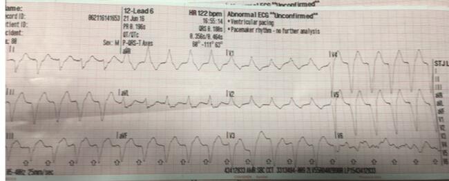
Tension pneumothorax (T-PTX) can precipitate rapid cardiopulmonary compromise and even death if not promptly identified and mitigated. While significant T-PTX is relatively uncommon, when it does occur, its presentation can be subtle and signs difficult to appreciate. A comprehensive review of the diagnosis of T-PTX is beyond the scope of this article, but it’s worth a brief mention that classically taught “hallmarks” like tracheal deviation and jugular venous distention are far less likely to be observed than are less specific signs such as tachycardia, hypoxia, respiratory distress, and ultimately, hypotension.1 Traditionally, treatment of a T-PTX in the prehospital setting has primarily included the use of needle thoracostomy (N-Th) as a mechanism to quickly vent the trapped gas that is compressing the lung and impeding intrathoracic blood flow.
Unfortunately, even when T-PTX is recognized, “darting the chest wall” is not always a simple task to perform correctly, and it’s not always easy to accomplish in a safe and effective manner. Therefore, those performing N-Th will need to appreciate the various limitations and certain clinical caveats for optimal performance, let alone the related potential for complications. Avoiding potential missteps and averting associated potential complications is pivotal to the success of this procedure. Most importantly, it will optimize the patient’s outcome under these time-critical circumstances.
Historically, the most common problem with N-Th is ineffective needle placement. Some studies have shown that, using traditional approaches, failure to penetrate the pleural space can occur in as many as 76% of attempted procedures and, accordingly, it does not decompress the life-threatening tension.1
This high rate of failure has since prompted two major changes to the technique now being used by many EMS systems. The first modification has been the use of longer needles compared to what was traditionally recommended. Previous recommendations involved the use of 5 cm (1.97 inch) needles. Longer needles, typically those 8 cm (3.25 inch) or more, are now the norm.2,3
The second modification was an update in N-Th site selection. Traditionally, rescuers were taught to go in over the top of the rib at the second or third intercostal space (ICS) in the mid-clavicular line of the involved lung. However, experience has shown that the needles can be misdirected using that proposed anatomical site.6,7
Therefore, in 2018, the 10th edition of the Advanced Trauma Life Support (ATLS) curriculum moved away from its recommendation for performing N-Th at the anterior chest and began endorsing a more lateral approach. Specifically, it was recommended to place the needle at the same site used for chest tube insertion, namely the fourth or fifth ICS near the anterior axillary line (AAL).4
That alternate location had several proposed advantages. The chest wall thickness may be thinner, depending on the patient’s body habitus, and thus it requires less distance to achieve penetration of the pleural cavity. In addition, the landmarks, specifically the ribs, can often be more easily palpated better ensuring proper placement above the rib.2,5 Furthermore, some might consider the lateral chest wall landmarks more easily identifiable than the anterior chest wall. Also, from a tactical medicine point of view, needle decompression can be achieved more rapidly in those individuals wearing body armor over the anterior chest.
The rationale for the move to the lateral site also had its roots in the documentation of complications at the anterior site. A growing body of literature had documented instances of improperly-placed needles and resulting complications including fatal penetration of mediastinal structures and great vessels using the anterior chest approach.8-10 With anterior placement, needles have sometimes been placed too far medially, leading to mediastinal and large vessel injury in some patients. In other cases, if the anterior insertion is too far superior, it can cause damage to the subclavian vasculature.13 Although a lateral approach may not eliminate the potential for iatrogenic injury entirely, it theoretically minimized these complications with respect to those vital structures. At the same time, the shift to a lateral approach with the longer needles may increase the potential of needle entrance into the left ventricle and other structures.
When needles are inserted too far inferiorly (occurring as often as 57% to 83% of the time), there is even potential penetration of the diaphragm, liver, stomach, small bowel, or spleen depending on placement side.10 Patient habitus, enlarged intra-abdominal contents (tense ascites, pregnancy, etc.), individual variations in pulmonary architecture, and aberrations in respiratory excursions may lead to increased risk at that lateral site. Also, in some cases, the anterior chest may be easier to palpate and penetrate compared to the lateral site.
Accordingly, the acknowledgement of these identified lateral approach complications has led to additional reassessment of more recent educational platforms for skills teaching, particularly those who still use only the fourth or fifth ICS at the level of the nipple or infra-mammary fold due to the variability in underlying anatomical structures.
The current thinking is that both sites are fraught with risk. Complications do occur in both locations, but both sets of complications can be avoided with improved anatomic knowledge. Accordingly, both the Tactical Combat Casualty Care and Tactical Emergency Casualty Care standards committees now indicate that either site can be reasonable for N-Th depending on all the above noted circumstances and understandings.
One relatively simple technique to aid in the rapid identification of a correct target site for lateral N-Th insertion is the use of a landmark-marking tool. The template is placed against the patient’s lateral chest with one end hooked into the armpit and the other pointing toward the hip, after which the area within a transparent window on the template identifies the third, fourth, or fifth ICS, specifically at the area between the mid- and anterior-axillary lines. Recognizing the inherent limitations of using a nonliving subject, a preliminary cadaver study showed a 96% success rate in identifying the appropriate landmarks for lateral N-Th, and a few larger studies of its efficacy are underway, hopefully ones that include dynamic physiologic and anatomic variances in live patients.12 How this compares to other guidance techniques such as placing the patient’s contralateral four fingers high in the axilla and using their intersection with the anterior aspect of the ipsilateral arm as the N-TH site is unresolved at this time. Additionally, with the increased use of ultrasound, this could become a guiding modality in prehospital N-Th, though more research would be needed, particularly in severely injured patients with variable types of respiratory excursions and organ locations.
Another potential evolution of the prehospital treatment of T-PTX is the “finger thoracostomy” procedure, which involves the use of a scalpel followed by blunt dissection to ventilate the pleural space in a manner similar to the initial steps for chest tube placement. This procedure brings with it the complexity of tools and techniques not as commonly used in EMS and an increased risk of provider injury, but it has the potential to decrease deep structure iatrogenic injury. However, it doesn’t necessarily address site selection precision. Additionally, use of this technique is currently limited in many places by EMS scope of practice regulations.
In summary, the treatment of T-PTX is a high-stress, time-dependent circumstance which may involve deep insertion of a large-bore needle into the chest cavity with potential risk of misplacement and iatrogenic injuries. Further affirmation of landmark devices, point-of-care ultrasound, and other procedures like finger thoracostomies, may guide approaches going forward. Current evidence supports the need for a broad understanding of the risks and benefits of different techniques and site selection as well as how patient body habitus and clinical scenario affect these.
References
1. Leigh-Smith S, Harris T. Tension prneumothorax - time for a re-think? Emerg Med J. 2005 Jan;22(1):8-16. doi:10.1136/emj.2003010421
2. Lesperance RN, Carroll CM, Aden JK, Young JC, Nunez TC: Failure rate of prehospital needle decompression for tension pneumothorax in trauma patients.(https://journals.sagepub.com/doi/10.1177/000313481808401130) Am Surg. 2018, 84:1750-5. 10.1177/000313481808401130
3. Inaba K, Branco BC, Eckstein M, et al. Optimal positioning for emergent needle thoracostomy: a cadaver-based study. Journal of Trauma. Nov 2011;71(5):1099-103; discussion 1103. doi:10.1097/TA.0b013e31822d9618
4. Chang SJ, Ross SW, Kiefer DJ, et al. Evaluation of 8.0-cm needle at the fourth anterior axillary line for needle chest decompression of tension pneumothorax. Journal of Trauma and Acute Care Surgery. 2014;76(4):1029-1034. doi:10.1097/ta.0000000000000158
5. Advanced trauma life support. 10th ed. American College of Surgeons; 2018.
6. Inaba K, Ives C, McClure K, et al. Radiologic evaluation of alternative sites for needle decompression of tension pneumothorax. Archives of Surgery. Sep 2012;147(9):813-8. doi:10.1001/archsurg.2012.751
7. Ferrie EP, Collum N, McGovern S. The right place in the right space? Awareness of site for needle thoracocentesis. Emergency Medicine Journal. 2005;22(11):788-789. doi:10.1136/emj.2004.015107
8. Lubin JS, Knapp J, Kettenmann ML. Paramedic Understanding of Tension Pneumothorax and Needle Thoracostomy (NT) Site Selection. Cureus. Jul 2022;14(7):e27013. doi:10.7759/cureus.27013
9. Wernick B, Hon HH, Mubang RN, et al. Complications of needle thoracostomy: A comprehensive clinical review. International Journal of Critical Illness and Injury Science. Jul-Sep 2015;5(3):160-9. doi:10.4103/2229-5151.164939
10. Axtman BC, Stewart K, Robbins JM, et al. Prehospital Needle Thoracostomy: What Are the Indications and Is a Post-Trauma Center Arrival Chest Tube Required? The American Journal of Surgery. 2019;doi:10.1016/j.amjsurg.2019.09.020
11. Thomas A, Wilkinson KH, Young K, Lenz T, Theobald J. Complications from Needle Thoracostomy: Penetration of the Myocardium. Prehospital Emergency Care. May-Jun 2021;25(3):438-440. doi:10.1080/10903127.2020.1772419
12. Fitzgerald M, Mackenzie CF, Marasco S, Hoyle R, Kossmann T. Pleural decompression and drainage during trauma reception and resuscitation. Injury. Jan 2008;39(1):9-20. doi:10.1016/j.injury.2007.07.021
13. Shah AN, Kothera CS, Dheer S. ThoraSite: A device to improve accuracy of lateral decompression needle and chest tube placement. J Trauma Acute Care Surg. Jul 2019;87(1S Suppl 1):S128-S131. doi:10.1097/TA.0000000000002244
14. Riwoe D, Poncia HD. Subclavian artery laceration: A serious complication of needle decompression. Emerg Med Australas. 2011 Oct;23(5):651-3. doi: 10.1111/j.1742-6723.2011.01466.x. PMID: 21995483.
About the Authors
Thomas W. Engel II, MD, MPH, is assistant professor in Emergency Medicine at the Medical College of Wisconsin.
Paul E. Pepe, MD, MPH, is is coordinator of the Metropolitan EMS Medical Director ("Eagles") Alliance, Dallas, Texas.
Dustin J. Calhoun, MD, FAEMS, is associate professor of emergency medicine at the University of Cincinnati College of Medicine.
Benjamin W. Weston, MD, MPH, FAEMS, is associate professor at the Medical College of Wisconsin and medical director for the Milwaukee County Office of Emergency Management.