Skin Color Assessment in EMS Education and Practice
Prehospital clinicians practice in an environment where time is short, conditions are uncontrolled, and assessment tools are limited. Because of this, EMS professionals are trained to rely on foundational physical exam findings—respiratory effort, mental status, skin signs, vital signs—to form rapid, accurate impressions of patient severity. Yet one of the most common elements of this initial assessment, skin color, is still taught using frameworks that do not reflect the diversity of patients encountered in modern EMS practice.
It is time to reconsider the role of skin color in prehospital medical decision-making and update the way we teach assessment to match scientific understanding, clinical evidence, and the reality of human biological variation.
Re-examining the 'General Impression'
At the beginning of every call, students are taught to form a general impression: Is the patient sick or not sick? Hurt or not hurt? Skin appearance is central to that impression. Traditional language—“pink, warm, and dry” for normal, and “pale, cool, and clammy” for shock—has been repeated so often that it has become assumed truth.
But is this accurate for all humans?
For patients with melanin-rich skin, “pink” is not a baseline. Pallor may not appear light or pale. Cyanosis may never manifest as blue. Teaching these descriptions as universal norms introduces systematic error into clinical reasoning, particularly during the crucial first minutes of patient contact, when EMS providers begin forming hypotheses about perfusion, oxygenation, and overall stability.
An updated approach should emphasize:
- Baseline skin tone varies widely across populations.
- Melanin can mask or delay visible color changes related to illness.
- Temperature and moisture often provide more reliable information than color alone.
- Assessment should include multiple anatomic locations—face, mucosa, nail beds, palms, and soles—to distinguish core from peripheral perfusion.
- Family or bystander input can help determine whether the patient’s current appearance is normal for them.
Changing documentation to “normal color, warm, and dry” rather than “pink, warm, and dry” is a small but meaningful step toward accuracy.
Why Teaching Resources Must Change
Existing EMS and medical textbooks often present rashes, cyanosis, hypoperfusion, dehydration, and anaphylaxis using images of light-skinned patients. This presents two problems:
- Students do not learn how conditions appear on diverse skin tones, making it harder for them to detect early signs in the field.
- Instructors—who were trained with the same limited images—may inadvertently perpetuate outdated assessment norms.
Dermatology research has documented these gaps. One study found that less than 19% of images in dermatology textbooks depict conditions on skin of color, and many physicians report inadequate training specific to patients with darker skin. EMS, which relies heavily on rapid visual cues, inherits this same structural limitation.
This has real clinical implications:
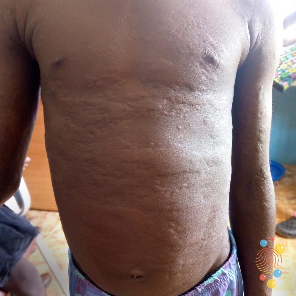
- Flushing may not appear red during anaphylaxis.
- Hives may be identifiable only by texture, not color.
- Shock may present as gray, ashen, or yellow-brown, rather than pale or cyanotic.
- Hyperthermia and hypothermia color changes may be subtle or delayed.
- Bruising may be difficult to identify early and requires palpation and symmetric comparison.
Updating images, case studies, and simulation scenarios is essential for safe clinical care.
Understanding Color Change in Melanin-Rich Skin
Melanin affects how light is absorbed and reflected by the skin, altering the visible manifestations of pathology. EMS providers should expect that:
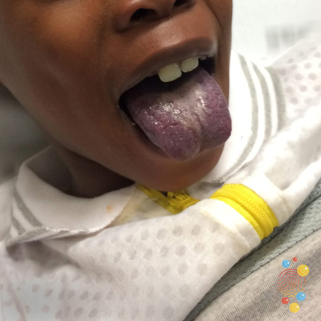
- Cyanosis is often a late—or absent—finding in darker skin tones.
- The most reliable locations to assess oxygenation changes are the lips, oral mucosa, nail beds, and conjunctiva rather than the skin surface.
- Ashen, gray, or dusky tones may represent hypoxia more accurately than “blue.”
- Peripheral cyanosis may present as darkening or coolness of fingertips rather than discoloration.
- In trauma, bruises may appear later because color contrast is less pronounced over melanin-rich skin.
Relying on “expected” textbook presentations can create dangerous blind spots. A patient without visible cyanosis may still be profoundly hypoxic. A trauma patient without visible ecchymosis may still have significant soft-tissue injury.
Pupillary Response: Another Layer of Complexity
Pupil assessment is another domain in which skin and eye pigmentation matter. Darker irises and reduced contrast between the iris and pupil can make it more difficult to evaluate reactivity, especially in low ambient light. EMS clinicians should:
- Use a brighter penlight or direct light source for pupils with less natural contrast.
- Assess in optimal lighting conditions whenever possible.
- Reassess multiple times if the environment is challenging.
These adjustments are simple but critical for accurate neurologic evaluation.
Pulse Oximetry: Technology Limitations That Impact Care
Pulse oximeters estimate oxygen saturation by measuring the absorption of red and infrared light through tissue. Because melanin also absorbs light, this technology is inherently more prone to error in patients with darker skin tones.
Recent research has shown that:
- Pulse oximeters may overestimate oxygen saturation in melanin-rich skin.
- These patients may receive less supplemental oxygen for the same true saturation.
- Device performance varies widely, and not all manufacturers test across skin tones.
For EMS practice, this means clinicians must:
- Interpret pulse oximetry cautiously, especially when the reading does not match the patient’s appearance, work of breathing, or mental status.
- Remember that pulse oximetry is an adjunct, not a substitute, for assessment.
- Advocate for devices validated across diverse skin tones as agencies upgrade equipment.
Technology should not undermine equitable care.
How We Got Here: A Brief History of 'Race' and Medicine
Modern racial categories based on skin color are a relatively recent invention—developed by European scientists over the last few centuries and intertwined with colonialism, slavery, and social hierarchy. These taxonomies were never biologically accurate. Today’s genomic research confirms:
- Human biological variation occurs on a spectrum, not in discrete racial categories.
- Variation within populations exceeds variation between populations.
- "Race" is a social and cultural construct, not a biological one.
Yet medical and dermatologic frameworks were built using these outdated classifications. As a result, conditions were described based on lighter-skinned populations, shaping everything from diagnostic criteria to textbook images to the common EMS teaching phrases we still use.
While race is not biologically meaningful, it is clinically relevant because it shapes:
- How people are treated,
- How their symptoms are perceived, and
- How accurately disease is recognized in them.
Prehospital medicine must address this gap because our patients depend on rapid, accurate interpretation of the signs in front of us.
Updating EMS Education: Moving Forward
Are we prepared to modernize the foundations of EMS education in light of these realities? We must be.
Initial training forms the base on which all clinical practice is built. When these foundations contain blind spots, they are reinforced across generations of providers and instructors. To prevent this:
1. Revise Textbooks and Curricula
- Replace “pink, warm, and dry” as a universal descriptor.
- Integrate examples of pathologic findings across a full spectrum of skin tones.
- Include evidence-based discussions of melanin’s impact on clinical assessment.
2. Diversify Teaching Images and Simulation Tools
- Use photographs, moulage kits, manikins, and case studies reflecting real-world diversity.
- Include scenarios demonstrating subtle or atypical presentations on darker skin.
3. Train for Tactile and Multisite Assessment
- Emphasize palpation, temperature, texture, and moisture.
- Teach students where color changes are most reliable on melanin-rich skin.
4. Update Clinical Documentation Practices
- Use inclusive, accurate terminology that aligns with modern science.
5. Advocate for Better Technology
- Support adoption of pulse oximeters validated for all skin tones.
- Educate clinicians on limitations until industry standards improve.
Ultimately, our mission in prehospital care is universal: to treat every patient at the highest standard possible, regardless of skin color, background, or presentation. Achieving that requires rethinking long-held assumptions and updating our educational structures to reflect the full diversity of the communities we serve.
Online Resources for Assessing Melanin-Rich Skin
- Black and Brown Skin – Mind the Gap: blackandbrownskin.co.uk/mindthegap
- American Nurse – Color Awareness in Patient Assessment: myamericannurse.com/color-awareness-a-must-for-patient-assessment/
- Brown Skin Matters (Instagram)
- Yale University Library – Skin of Color Resource List: guides.library.yale.edu/skin-of-color
- Lilly – Skin of Color Education: skinofcolor.lilly.com
About the Authors
Justin S. Padgett, M.S., Paramedic, WEMT, and NCOEMS Program Coordinator, is the Executive Director of Landmark Learning. Since 1996, Landmark Learning has been dedicated to providing students with training in out-of-hospital care, wilderness medicine, and water rescue. The Landmark Learning campus in Cullowhee, NC, trains over 3,000 students annually. As the Curriculum Director at Landmark, Justin has spent the past 29 years training instructors to educate students effectively. His experience in out-of-hospital care, wilderness medicine, travel medicine programs, and adventure travel provides him with a unique perspective on achieving tangible results. He believes that preparing students for real-world scenarios requires instructors to blend experience, validity, andragogy, and immersive education.
Seth Collings Hawkins, MD, MA, MPH, is an anthropologist-physician, double board-certified in emergency medicine and EMS. He’s the first physician named Master Fellow of the Academy of Wilderness Medicine. He’s also a Fellow of the Academy of EMS, the American College of Emergency Physicians, and the American Academy of Emergency Medicine. He’s an Associate Professor of Emergency Medicine at Wake Forest University and Associate Director of its Wilderness Medicine Fellowship. He serves as medical director for Landmark Learning, the National Association for Search & Rescue, and NC State Parks, and as LEMA for all US Forests in NC (USDA Forest Service) and the Outer Banks (National Park Service).